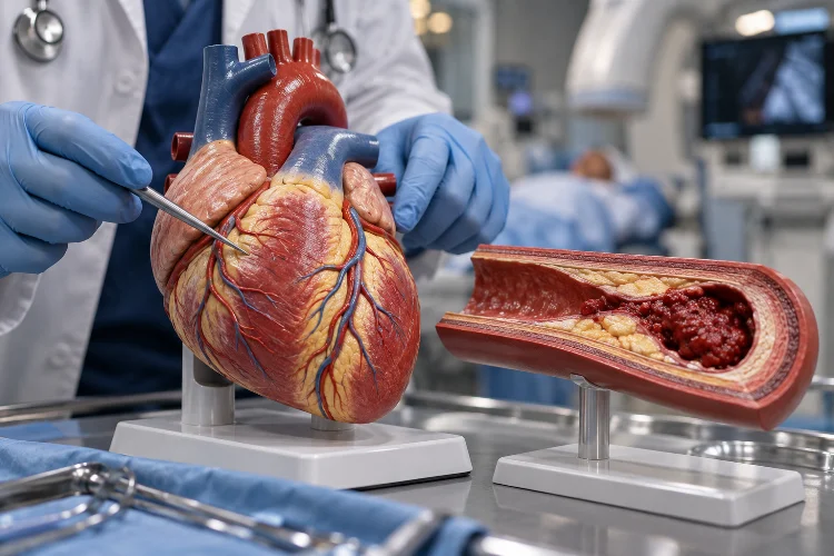
A heart attack may feel like a sudden event, but the biological story behind it often begins years earlier.
Slow changes inside the coronary arteries can gradually reduce blood flow to the heart. Then, sometimes within minutes, a disrupted plaque and newly formed blood clot can transform a relatively stable condition into a medical emergency.
Understanding the pathology of heart attacks and ischemia helps explain what is actually happening during this process. Ischemia means that heart tissue is not receiving enough oxygen-rich blood.
If the shortage is severe or lasts too long, cardiac muscle cells become injured and eventually begin to die, producing what doctors call a myocardial infarction.
The process connects several major pathology concepts: atherosclerosis, oxygen deprivation, cellular injury, necrosis, inflammation, healing, and scar formation.
Once you follow this sequence, terms such as myocardial ischemia, STEMI, NSTEMI, and cardiac troponin become much easier to understand. More importantly, you can see why restoring blood flow quickly is so critical during a heart attack.
What Is Myocardial Ischemia?
Ischemia occurs when blood flow to a tissue becomes inadequate for its metabolic needs. In myocardial ischemia, the tissue affected is the heart muscle, or myocardium.
Because cardiac muscle works continuously, it requires a reliable supply of oxygen and nutrients.
When coronary blood flow falls, oxygen delivery may no longer match the heart’s demand. The American Heart Association describes cardiac ischemia as decreased blood flow and oxygen to the heart muscle.
Mild or temporary ischemia does not always cause permanent cell death. A person may experience angina during physical activity because the heart needs more oxygen than a narrowed coronary artery can deliver.
Ischemia can even occur without obvious symptoms, a condition known as silent ischemia.
This distinction matters: ischemia describes insufficient blood supply, while infarction means that prolonged ischemia has caused irreversible tissue death.
How Atherosclerosis Sets the Stage for a Heart Attack
Most heart attacks are connected to coronary artery disease, in which atherosclerotic plaque develops inside the walls of the coronary arteries. Plaque contains substances such as cholesterol, fat, calcium, and cellular material.
As plaque accumulates, the artery may gradually become narrower. This can reduce the amount of oxygen-rich blood reaching cardiac muscle, particularly when oxygen demand increases during exercise or stress.
However, severe narrowing is not the only danger.
An atherosclerotic plaque can become unstable and rupture or erode. When that happens, substances inside the plaque are exposed to circulating blood. Platelets become activated, the coagulation system responds, and a thrombus – or blood clot – can rapidly form.
That clot may partially or completely block a coronary artery.
This explains why someone can appear relatively stable and then suddenly develop an acute coronary syndrome. The long-term disease and the sudden event are seperate parts of the same pathological story.
What Happens to Heart Cells During Ischemia?
Once coronary blood flow drops significantly, cardiac cells begin experiencing oxygen deprivation.
Normally, heart muscle cells use oxygen to generate the energy needed for contraction, membrane function, and other cellular activities. With inadequate oxygen, energy production becomes less efficient and normal cellular processes start to fail.
The myocardium may initially lose some of its ability to contract and relax properly. Ischemic areas can therefore become weak or move abnormally even before permanent tissue death has occured.
If normal blood flow returns early enough, some of these changes can be reversible.
Persistent ischemia is different. Cell membranes, mitochondria, proteins, and other structures become progressively damaged. NCBI’s review of myocardial infarction notes that prolonged deprivation of oxygen ultimately causes myocardial cell death and necrosis.
This transition from reversible injury to irreversible injury is one of the most important moments in heart attack pathology.
When Ischemia Becomes a Myocardial Infarction
A myocardial infarction, commonly called a heart attack, occurs when part of the myocardium develops necrosis in the setting of myocardial ischemia.
The amount of damage depends on several factors, including how completely blood flow is blocked, which coronary artery is involved, how much myocardium it supplies, and how long severe ischemia continues.
The CDC emphasizes that the longer treatment is delayed, the greater the potential damage to heart muscle.
One useful pathological concept is that necrosis does not necessarily destroy the entire affected region at exactly the same moment.
The inner portion of the ventricular wall, called the subendocardium, is especially vulnerable to ischemia. With prolonged severe obstruction, injury can extend outward through more of the myocardial wall.
This is one reason the phrase “time is muscle” is so meaningful in emergency cardiology. Restoring blood flow quickly may preserve myocardium that is ischemic but not yet irreversibly damaged.
STEMI and NSTEMI: What Is the Difference?
Two terms health students quickly encounter are STEMI and NSTEMI.
STEMI means ST-segment elevation myocardial infarction. NSTEMI means non-ST-segment elevation myocardial infarction. These classifications are primarily based on ECG findings together with clinical and laboratory evidence rather than simply the microscopic depth of tissue death.
A STEMI often results from sustained severe or complete coronary obstruction and is associated with significant myocardial injury. Because rapid reperfusion can save endangered heart tissue, STEMI commonly requires urgent restoration of coronary blood flow.
NSTEMI also involves myocardial necrosis, but the characteristic acute ST-segment elevation is absent. The obstruction may be incomplete or involve different patterns of ischemia and injury.
There is also unstable angina, which belongs to the acute coronary syndrome spectrum but traditionally involves ischemia without detectable myocardial necrosis.
Modern high-sensitivity troponin testing has helped clinicians identify small amounts of myocardial injury that might previously have gone undetected.
Why Troponin and ECG Changes Matter
The pathology of myocardial infarction is happening inside heart tissue, so clinicians need ways to detect it without directly examining the myocardium under a microscope.
One major tool is the electrocardiogram (ECG). Ischemic and injured cardiac cells behave differently electrically, producing changes such as ST-segment elevation, ST depression, or T-wave abnormalities depending on the situation.
Another important tool is cardiac troponin.
Troponins are proteins associated with cardiac muscle cells. When those cells are damaged, troponin can enter the bloodstream.
A characteristic rise and/or fall in cardiac troponin, interpreted together with evidence of myocardial ischemia, is central to diagnosing myocardial infarction.
This is a good example of pathology connecting microscopic injury with measurable clinical evidence.
The damaged cell cannot simply tell a doctor what happened, but its released proteins provide biochemical clues.
How a Heart Attack Affects Heart Function
A heart attack does more than destroy individual cardiac cells.
Heart muscle needs enough strenght to pump blood throughout the body. When a significant section becomes ischemic or necrotic, that region may contract poorly or stop contracting normally.
If enough myocardium is damaged, cardiac output can fall. Patients may develop heart failure, low blood pressure, pulmonary congestion, or, in severe cases, cardiogenic shock.
Electrical instability is another important complication.
Damaged and ischemic cardiac cells may conduct electrical signals abnormally, increasing the risk of arrhythmias. Some dangerous ventricular arrhythmias can prevent the heart from pumping effectively and contribute to sudden cardiac death.
Depending on the location of the infarction, damage may also affect papillary muscles, the interventricular septum, or other important structures. Mechanical complications are less common but can be extremely serious.
This is why infarct size and location matter, not simply whether a heart attack happened.
What Happens After the Heart Muscle Dies?
Unlike some tissues, adult cardiac muscle has very limited ability to regenerate after major necrotic injury.
After an infarction, the body begins clearing damaged tissue and initiating repair. Inflammatory cells participate in removing dead cellular material, while healing processes eventually replace the lost myocardium with fibrous scar tissue.
Scar tissue can help maintain structural integrity, but it does not contract like healthy cardiac muscle.
As a result, a large infarction can permanently alter the way the heart pumps. The remaining myocardium may also undergo structural changes known as ventricular remodeling, including changes in chamber size, shape, and wall characteristics.
Over time, these changes may contribute to chronic heart failure or other cardiovascular problems.
The pathology therefore continues long after the original coronary blockage has been treated.
Why Rapid Treatment Changes the Pathological Outcome
Heart attack treatment is closely connected to the biology of ischemic injury.
If an obstructed coronary artery is reopened before too much myocardium dies, blood and oxygen can once again reach threatened tissue. This process is called reperfusion.
Percutaneous coronary intervention, commonly involving angioplasty and stent placement, is one major method of restoring blood flow. In some circumstances, fibrinolytic medication may also be used for STEMI when timely PCI is unavailable.
The goal is not merely to make chest pain disappear. The deeper goal is to prevent ischemic tissue from progressing to irreversible necrosis.
Anyone experiencing possible heart attack symptoms – such as persistent chest pressure or discomfort, shortness of breath, pain spreading to the arm, jaw, neck, or back, cold sweating, weakness, or unexplained nausea – should seek emergency medical attention immediately.
The sooner patients recieve appropriate treatment, the more heart muscle may be preserved.
Understanding the pathology of heart attacks and ischemia turns a familiar medical emergency into a clear biological sequence. Coronary artery disease can gradually develop over many years, while plaque disruption and thrombosis may suddenly reduce blood flow.
Oxygen deprivation then causes myocardial ischemia, cellular dysfunction, irreversible injury, and eventually necrosis if circulation is not restored.
The resulting damage can affect pumping ability, electrical conduction, and long-term cardiac structure. This is also why ECG findings, cardiac troponin, and rapid reperfusion are so important in clinical care.
For health students, mastering this process creates a strong foundation for learning cardiovascular disease. Next, explore atherosclerosis, acute coronary syndromes, cardiac biomarkers, and post-infarction healing to connect the pathology with diagnosis and treatment.