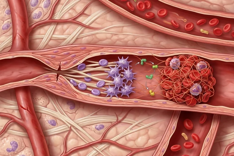
A tiny cut can damage dozens of blood vessels, yet most minor injuries stop bleeding within minutes. That quick response is possible because the body has a highly coordinated system called haemostasis.
Normal haemostasis stops blood loss at the site of vascular injury while keeping blood flowing normally everywhere else. It is not simply “blood clotting.”
The process involves the vessel wall, platelets, coagulation proteins, natural anticoagulant mechanisms, and eventually fibrinolysis, which helps remove the clot when it is no longer needed.
Understanding how normal haemostasis stops bleeding after vascular injury is essential for medical learners because problems anywhere along this pathway can produce either excessive bleeding or unwanted thrombosis.
The process is easiest to understand as a sequence:
Vascular injury → vasoconstriction → platelet adhesion → platelet activation and aggregation → thrombin generation → fibrin formation → stable clot → controlled clot removal
Each stage overlaps with the next, creating a fast but tightly regulated response.
What Is Normal Haemostasis?
Haemostasis is the physiological process that limits blood loss following damage to a blood vessel while preserving normal circulation elsewhere.
This balancing act is crucial. If the response is too weak, even minor injuries may lead to prolonged bleeding. If it becomes excessive or spreads beyond the damaged area, an unwanted thrombus may obstruct normal blood flow.
Haemostasis is commonly divided into several connected components.
Primary haemostasis mainly involves the injured vessel and platelets forming an initial platelet plug.
Secondary haemostasis strengthens that plug by generating fibrin through the coagulation system. Later, regulatory mechanisms restrict clot formation, while fibrinolysis gradually removes fibrin as healing proceeds.
These are not completely separate events. Platelets and coagulation proteins communicate constantly during clot formation.
Vascular Injury Immediately Changes the Vessel Wall
The first event is damage to the vascular endothelium.
Healthy endothelial cells normally provide a surface that discourages unnecessary platelet activation and coagulation.
Once the endothelial layer is disrupted, blood becomes exposed to structures underneath it, including extracellular matrix proteins that normally remain hidden from circulating platelets.
The damaged blood vessel also undergoes vasoconstriction.
Narrowing the vessel temporarily reduces blood flow through the injured area. This does not permanently stop bleeding on its own, but it gives the other components of haemostasis time to respond.
You can think of vasoconstriction as the body’s first emergency measure:
Make the hole smaller and slow the blood while the repair team arrives.
Platelets then provide the next major response.
Platelets Stick to the Damaged Vessel
Platelets normally circulate without attaching strongly to healthy vascular surfaces.
When the endothelial lining breaks, however, subendothelial components become exposed.
Platelets begin adhering to the injured area, with von Willebrand factor (vWF) playing an important role in connecting exposed vessel-wall structures with platelet receptors, particularly under flowing blood conditions.
Primary haemostasis involves vasoconstriction followed by platelet adhesion, activation, and aggregation.
Platelet Adhesion Anchors the First Responders
Platelet adhesion means circulating platelets attach to the damaged surface rather than simply flowing past it.
This initial attachment is crucial because it concentrates platelets exactly where the vessel has been broken.
A useful analogy is patching a leaking pipe. Before you can build a strong repair, something first needs to grip the damaged edge.
That is what platelet adhesion accomplishes.
But attached platelets do not remain quiet. Contact with the injured surface activates them.
Activated Platelets Build the Primary Platelet Plug
Platelet activation produces major changes in platelet shape and function.
Activated platelets become better able to interact with one another and release chemical mediators that recruit additional platelets. They also provide membrane surfaces on which important coagulation reactions can occur.
One important mediator released during platelet activation is ADP, which helps recruit and activate additional platelets. Platelets also generate thromboxane A2, which contributes to further platelet activation and vascular responses.
Activated platelets expose functional glycoprotein IIb/IIIa receptors, which allow fibrinogen to bridge neighbouring platelets.
This creates platelet aggregation.
More platelets join, bind together, and form a temporary structure over the injured vessel.
The sequence is therefore:
Adhesion → activation → recruitment → aggregation → platelet plug
This plug can rapidly reduce blood loss, but at this stage it is still relatively fragile.
It needs reinforcement.
The Coagulation System Produces Thrombin
Secondary haemostasis strengthens the platelet plug through activation of the coagulation system.
Coagulation involves a series of plasma proteins that circulate largely as inactive precursors. After vascular injury, these factors become activated in a controlled sequence that ultimately produces the enzyme thrombin.
Traditional teaching divides coagulation into intrinsic, extrinsic, and common pathways. This framework remains useful for understanding laboratory tests, although coagulation inside the body is better viewed as a highly interconnected process occurring on cellular surfaces.
Tissue factor exposed at an injury site is particularly important for initiating coagulation in vivo.
As coagulation reactions progress, activated factor X participates in converting prothrombin into thrombin.
Thrombin then becomes one of the central molecules in haemostasis.
It amplifies coagulation, activates additional platelets, and most importantly converts soluble fibrinogen into fibrin.
Fibrin Turns the Platelet Plug Into a Stable Clot
A platelet plug works like a temporary patch. Fibrin provides reinforcement.
Thrombin cleaves fibrinogen molecules, allowing fibrin molecules to polymerize into long strands. These strands form a network around the aggregated platelets.
The fibrin mesh traps additional cells and strengthens the developing haemostatic plug.
Factor XIII helps cross-link fibrin, making the network more mechanically stable.
The result is a stable platelet-fibrin clot capable of resisting the pressure of flowing blood while the damaged vessel begins healing.
The difference between primary and secondary haemostasis can therefore be remembered simply:
Primary haemostasis builds the platelet plug.
Secondary haemostasis reinforces it with fibrin.
The two systems work closely together rather than acting independently. Platelets provide surfaces needed for efficient coagulation reactions, while thrombin generated through coagulation further activates platelets.
Why Clotting Does Not Spread Through the Entire Circulation
Once you understand how rapidly coagulation can amplify, an obvious question appears:
Why doesn’t one small injury cause the entire bloodstream to clot?
Normal haemostasis includes powerful regulatory mechanisms that keep coagulation localized.
Healthy surrounding endothelial cells remain antithrombotic. Blood flow dilutes and removes activated coagulation factors, while endogenous anticoagulant pathways restrain excessive thrombin generation.
Important regulatory systems include antithrombin, the protein C system, and tissue factor pathway inhibitor.
These mechanisms allow the body to build a clot where the vessel is damaged while maintaining fluid blood elsewhere. Physiological haemostasis therefore depends just as much on limiting clot formation as it does on starting it.
This balance can be summarized as:
Injury site → pro-clotting
Healthy circulation → anti-clotting
Losing that balance can lead toward either haemorrhage or thrombosis.
What Happens After the Bleeding Has Stopped?
A clot is useful during vessel repair, but it should not remain indefinitely.
As the vessel heals, the haemostatic plug is gradually reorganized and eventually removed through fibrinolysis.
The central enzyme in fibrinolysis is plasmin.
Plasmin is generated from an inactive precursor called plasminogen and breaks fibrin into smaller degradation products. This allows the fibrin network to disappear as tissue integrity is restored.
Haemostasis therefore includes both clot formation and appropriate clot removal. The American Society of Hematology recognizes platelet plug formation, fibrin clot formation, and fibrinolysis as major functional phases of the haemostatic system.
Fibrinolysis also has to be controlled.
Removing the clot too early could restart bleeding, while inadequate fibrinolysis could allow unnecessary clot material to persist.
Normal haemostasis is therefore about timing and location, not merely clot production.
Primary and Secondary Haemostasis Explain Different Bleeding Patterns
The distinction between platelet function and coagulation becomes clinically useful when haemostasis fails.
Disorders involving platelets or von Willebrand factor commonly interfere with primary haemostasis. These problems often produce mucosal bleeding, easy bruising, nosebleeds, or prolonged bleeding from small injuries.
Deficiencies of coagulation factors interfere more strongly with secondary haemostasis.
Haemophilia is a classic example. The initial platelet plug can form, but insufficient fibrin reinforcement makes durable haemostasis more difficult.
The American Society of Hematology explains that normal clotting requires both platelets forming a plug and coagulation proteins generating fibrin that holds that plug securely in place.
This provides a useful clinical lesson:
Platelets create the first seal; fibrin makes the seal last.
A Simple Way to Remember Normal Haemostasis
For medical learners, the pathway can be reduced to six steps:
Constrict → Stick → Activate → Aggregate → Reinforce → Remove
First, the injured blood vessel constricts.
Platelets then stick to the exposed vessel wall and become activated. Additional platelets arrive and aggregate, creating the primary haemostatic plug.
The coagulation system generates thrombin, which converts fibrinogen into fibrin. Fibrin then reinforces the platelet plug and creates a stable clot.
Finally, after healing progresses, fibrinolysis helps remove the fibrin network.
Another useful sequence is:
Vessel wall → Platelets → Thrombin → Fibrin → Stable clot → Fibrinolysis
If you can explain why each step leads to the next, you already understand the basic logic of normal haemostasis.
Normal haemostasis stops bleeding after vascular injury through a carefully controlled partnership between blood vessels, platelets, coagulation proteins, fibrin, anticoagulant mechanisms, and fibrinolysis.
Vasoconstriction first reduces blood flow. Platelets adhere to the damaged surface, become activated, and aggregate into a primary plug. Coagulation then generates thrombin, which converts fibrinogen into fibrin and reinforces the plug into a stable clot.
Just as importantly, natural anticoagulant mechanisms restrict the response to the injured area, while fibrinolysis removes the clot as healing progresses.
For medical learners, remember the sequence rather than memorizing every factor first: slow the bleeding, build the platelet plug, strengthen it with fibrin, control its spread, and remove it after repair.
Use that framework when you next study haemophilia, von Willebrand disease, thrombocytopenia, or thrombosis.