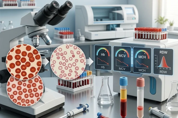
A complete blood count may show low hemoglobin, but that number alone does not tell the whole story.
To understand what is happening in anemia, medical learners need to look at the red blood cells themselves: their size, hemoglobin content, and how much they vary from one cell to another.
Iron deficiency creates a particularly recognizable pattern.
Iron is essential for making hemoglobin, the protein inside red blood cells that carries oxygen. When iron becomes scarce, the bone marrow gradually loses its ability to produce normally hemoglobinized erythrocytes.
The resulting cells eventually become smaller and paler, producing the classic microcytic, hypochromic anemia associated with iron deficiency.
Understanding how iron deficiency changes red cells and their indices is useful because the CBC often reveals the biological process before you even look at a blood smear.
The key sequence is straightforward: iron stores fall → hemoglobin production decreases → new red cells become smaller and paler → MCV and MCH fall → RDW often rises.
Why Red Blood Cells Need Iron
Red blood cells exist mainly to transport oxygen, and hemoglobin makes that possible.
Iron sits at the center of the heme component of hemoglobin. Without enough available iron, developing erythroid cells in the bone marrow cannot produce normal amounts of hemoglobin.
MedlinePlus notes that iron is required to make hemoglobin, which carries oxygen from the lungs to tissues throughout the body.
At first, the body can draw on stored iron.
As those reserves become depleted, less iron is available for erythropoiesis. Ferritin generally falls as iron stores decline, followed by reduced circulating iron availability and transferrin saturation. Eventually, hemoglobin production becomes impaired enough to produce anemia.
This explains why iron deficiency can exist before anemia appears.
A person may have depleted iron stores while hemoglobin and red cell indices are still within the laboratory reference range. The CBC becomes more obviously abnormal as the deficiency progresses.
MCV Falls as Red Cells Become Microcytic
One of the most familiar red cell indices is mean corpuscular volume, or MCV.
MCV represents the average volume, or size, of circulating red blood cells. When the average cell becomes smaller than the laboratory reference range, the anemia is described as microcytic.
Iron deficiency is one of the most common causes of microcytic anemia.
Why do the cells become smaller?
Developing erythrocytes need to accumulate enough hemoglobin during maturation. When hemoglobin synthesis is inadequate, the developing cells undergo additional divisions before reaching their final stage, producing smaller mature red cells.
The important point is that MCV does not necessarily fall immediately.
Early iron deficiency may still appear normocytic, especially before iron restriction becomes severe enough to significantly alter new red cell production. As deficiency progresses, microcytosis becomes more obvious.
For learners, remember:
Early iron deficiency → MCV may still be normal
Established iron deficiency anemia → MCV commonly becomes low
MCH Falls Because Each Cell Contains Less Hemoglobin
Another useful CBC value is mean corpuscular hemoglobin, or MCH.
MCH estimates the average amount of hemoglobin contained in each red blood cell. Because iron deficiency interferes with hemoglobin synthesis, newly produced cells contain less hemoglobin, so MCH tends to decrease.
This is closely related to microcytosis.
Smaller cells generally contain less total hemoglobin, which is why MCH often falls alongside MCV. Classical descriptions of iron deficiency anemia therefore refer to the cells as microcytic and hypochromic.
Under the microscope, hypochromic red cells show an enlarged pale area in the center because there is less hemoglobin filling the cell.
Normally, red blood cells have some central pallor because of their biconcave shape. In established iron deficiency, that pale region becomes more prominent.
So the laboratory value and the microscope appearance tell the same biological story:
Less iron → less hemoglobin → lower MCH → increased central pallor
What Happens to MCHC?
Mean corpuscular hemoglobin concentration, or MCHC, represents the average concentration of hemoglobin within the red blood cell volume.
Students often expect MCHC to fall dramatically in every case of iron deficiency because the cells are called hypochromic. In reality, MCHC can be less sensitive than MCV or MCH and may remain within the normal range, particularly in less advanced disease.
Older and modern automated measurements do not always classify microcytic anemia as strongly hypochromic based on MCHC alone.
With more established iron deficiency, however, reduced hemoglobinization may produce a low MCHC.
This is why no single red cell index should be interpreted by itself.
If the hemoglobin is low, MCV and MCH are reduced, RDW is increased, and the smear shows microcytic hypochromic cells, the pattern becomes much more informative than any one number.
RDW Often Rises as Cell Sizes Become Unequal
Red cell distribution width, or RDW, tells you how much red blood cell size varies across the circulating population.
When cells are fairly similar in size, RDW tends to remain relatively narrow. When there is a wider mixture of large and small cells, RDW rises.
This variation is called anisocytosis.
Iron deficiency commonly produces an elevated RDW because the disease develops over time. Older circulating red cells may have been produced when iron availability was better, while newer cells become progressively smaller as deficiency worsens.
The bloodstream therefore contains a mixture of cell sizes.
This makes RDW particularly helpful when interpreted together with MCV. Iron deficiency classically produces low MCV with increased RDW, although the exact pattern depends on disease stage and other coexisting conditions.
That contrasts with some thalassemia traits, where marked microcytosis may occur with a more uniform red cell population and therefore a less elevated RDW. The distinction is useful as a clue, but it is not diagnostic by itself.
What the Peripheral Blood Smear Looks Like
The automated CBC gives numerical clues, but a peripheral blood smear lets you see the consequences directly.
In established iron deficiency anemia, red cells typically appear microcytic and hypochromic. The central area of pallor becomes larger, reflecting reduced hemoglobin content.
Variability in both size and shape may also increase.
The terms anisocytosis and poikilocytosis describe variation in cell size and abnormal cell shapes, respectively. Iron deficiency can produce anisopoikilocytosis, and elongated elliptocytes sometimes described as pencil cells may appear in more established disease.
The smear therefore adds a visual layer to the CBC:
Low MCV → small cells
Low MCH → less hemoglobin per cell
High RDW → greater variation in cell size
Blood smear → microcytosis, hypochromia, and variable shapes
Once those connections become familiar, red cell indices stop feeling like unrelated laboratory abbreviations.
How the CBC Pattern Changes as Iron Deficiency Progresses
Iron deficiency develops in stages rather than instantly.
Early on, stored iron becomes depleted. Ferritin may become abnormal before hemoglobin falls significantly. Red cells already circulating in the bloodstream may still look relatively normal because they were produced when iron availability was better.
As deficiency continues, the bone marrow receives less usable iron.
Hemoglobin synthesis falls, newer red cells become less well hemoglobinized, and RDW may increase as these smaller cells mix with older normal-sized erythrocytes.
Eventually, the classic pattern becomes more obvious:
Hemoglobin ↓
MCV ↓
MCH ↓
RDW ↑
Peripheral smear → microcytic, hypochromic cells with anisopoikilocytosis
MCHC may decrease as well, particularly in more pronounced hypochromia, but it can be less consistently abnormal than MCV and MCH.
This progression explains why a normal MCV does not automatically exclude early iron deficiency.
Red Cell Indices Suggest Iron Deficiency but Do Not Confirm It
A microcytic CBC pattern is strongly suggestive, but iron deficiency is not the only cause.
Thalassemia, anemia of inflammation, sideroblastic disorders, and lead-related abnormalities can also produce microcytosis under certain circumstances.
That is why iron studies are important.
Serum ferritin is widely used to assess stored iron, while transferrin saturation helps estimate how much circulating iron is available for tissues. Ferritin is particularly useful, although inflammation can raise ferritin and make interpretation more complicated.
A clinician therefore does not diagnose iron deficiency from MCV alone.
The more complete approach is:
CBC pattern + peripheral smear + ferritin + transferrin saturation + clinical context
The underlying cause also matters. Iron deficiency may develop because of chronic blood loss, inadequate iron intake, increased requirements, or impaired absorption, so identifying the deficiency should usually lead to another question: Why is the patient losing or not obtaining enough iron?
A Simple Way to Read Red Cell Indices
For medical learners, begin with hemoglobin.
If it is low, look at MCV to decide whether the anemia is microcytic, normocytic, or macrocytic.
If MCV is low, look at MCH and RDW. Low MCH supports reduced hemoglobinization, while increased RDW suggests greater variation in red cell size.
Then check the blood smear and iron studies.
A classic iron deficiency pattern can be summarized as:
Iron stores fall → hemoglobin synthesis falls → cells become smaller → cells contain less hemoglobin → cell-size variation increases
Or, in laboratory language:
Ferritin ↓ → Hb ↓ → MCV ↓ → MCH ↓ → RDW ↑
The exact order and degree of abnormality vary, so the pattern should always be interpreted as a whole rather than as a rigid formula.
Iron deficiency changes red cells and their indices because developing erythrocytes do not have enough iron to make normal amounts of hemoglobin.
As deficiency progresses, the cells become smaller and less well hemoglobinized, producing the classic microcytic, hypochromic appearance of iron deficiency anemia.
On the CBC, this commonly appears as falling hemoglobin, low MCV, low MCH, and increased RDW. A peripheral smear may reveal microcytosis, increased central pallor, anisocytosis, and abnormal cell shapes.
For medical learners, do not memorize the indices separately. Follow the biology: less iron means less hemoglobin, less hemoglobin produces smaller paler cells, and progressive deficiency creates a mixed population of red cell sizes.
Next time you see a CBC, try reading the red cell indices as a story rather than a set of numbers.