A routine blood test can tell you that a patient’s white blood cell count is unusually high. But it cannot always explain why.
Is the increase caused by infection, inflammation, leukemia, or another bone marrow disorder? Answering that question often requires a closer look at the cells themselves.
That is where hematopathology comes in.
So, what is hematopathology? It is a specialized field of pathology focused on diseases involving blood-forming cells, bone marrow, lymphoid tissues, and related hematologic systems.
The College of American Pathologists defines hematopathology as the study and diagnosis of diseases involving hematolymphoid cells and tissues as well as blood coagulation.
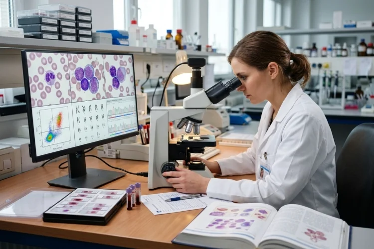
Hematopathologists do much more than look at blood under a microscope. They combine cell morphology with information from bone marrow biopsies, lymph nodes, flow cytometry, immunohistochemistry, chromosome studies, and molecular tests.
For medical learners, hematopathology becomes much easier when viewed as a detective process: find the abnormal cells, identify their lineage, understand their genetics, and connect everything with the clinical picture.
What Does Hematopathology Study?
Hematopathology sits at the intersection of pathology and hematology.
The field deals with disorders involving hematopoietic cells, which give rise to red blood cells, white blood cells, and platelets, as well as lymphoid tissues such as lymph nodes.
Hematopathologists may evaluate blood, bone marrow, lymph nodes, spleen specimens, and other tissues where hematologic disease is suspected. CAP notes that the specialty combines evaluation of blood, cellular phenotype and morphology, coagulation results, and bone marrow biopsies.
Many of the diseases encountered are cancers, including leukemias, lymphomas, plasma cell neoplasms, myelodysplastic neoplasms, and myeloproliferative neoplasms.
However, hematopathology is not simply “cancer pathology.” Abnormal blood-cell production and non-neoplastic hematologic conditions can also produce important morphological findings that require careful investigation.
The challenge is that many disorders can look similar at first.
A high white blood cell count, for example, can occur during infection or leukemia. The number itself is only the beginning of the diagnostic story.
The Peripheral Blood Smear Provides the First Visual Clues
One of the simplest but most powerful tools in hematopathology is the peripheral blood smear.
A small drop of blood is spread across a glass slide, stained, and examined under a microscope. This allows the examiner to directly assess the size, shape, maturity, and appearance of blood cells.
MedlinePlus notes that blood smears provide information about the number and morphology of red cells, white cells, and platelets and can reveal abnormalities associated with leukemia, anemia, and bone marrow disorders.
Imagine a patient with severe leukocytosis.
The complete blood count tells you that white cells are increased. The smear provides another level of information: Are those cells mostly mature neutrophils, reactive lymphocytes, or immature blasts?
Blasts are immature hematopoietic precursor cells that normally live primarily in the bone marrow. Finding significant numbers in peripheral blood can raise concern for an acute leukemia, although morphology alone does not always establish the exact type.
This is a recurring principle in hematopathology:
Numbers identify the abnormality; morphology begins explaining it.
Bone Marrow Shows Where Blood Cells Are Being Made
Sometimes peripheral blood provides enough information to strongly suspect a diagnosis. In many cases, however, hematopathologists need to examine the bone marrow, where blood-cell production takes place.
Bone marrow testing commonly involves two related procedures.
A bone marrow aspiration removes liquid marrow containing individual cells. A bone marrow core biopsy removes a small piece of bone and marrow, preserving the overall tissue architecture.
These tests can help diagnose disorders such as leukemia, lymphoma involving the marrow, myelodysplastic neoplasms, and other hematologic diseases.
The aspiration is useful for examining individual cells in detail.
The core biopsy answers different questions. Is the marrow unusually crowded? Has normal blood production been replaced by malignant cells? Is fibrosis present? Are abnormal cells distributed diffusely or in clusters?
For example, in acute leukemia, a bone marrow specimen may show replacement of normal hematopoietic tissue by blasts. Blood and bone marrow examinations are major components of the diagnostic workup for acute myeloid leukemia.
Together, the aspirate and biopsy provide both cell-level and tissue-level information.
Flow Cytometry Identifies a Cell’s Immunophenotype
Cells may look alike under a microscope while actually belonging to different biological lineages.
That is why flow cytometry is so important in modern hematopathology.
Flow cytometry analyzes individual cells using markers recognized by fluorescently labeled antibodies.
The cells pass through a laser-based system, allowing multiple surface or intracellular proteins to be measured. This creates an immunophenotype-essentially a molecular identity card for the cell population.
Suppose a marrow contains many blasts.
Morphology tells the hematopathologist that an acute leukemia may be present, but treatment depends heavily on whether the cells belong to a myeloid, B-lymphoid, T-lymphoid, or less common lineage.
Flow cytometry can answer that question by examining patterns of markers such as CD19, CD3, CD13, CD33, CD34, and many others.
In acute myeloid leukemia, NCI guidance emphasizes that immunophenotyping by flow cytometry plays an important role in determining leukemic cell lineage because morphology alone may be insufficient.
So the workflow becomes:
Abnormal cells → morphology → immunophenotype → more precise classification
Lymph Node Biopsies Help Diagnose Lymphoma
Leukemia often has major involvement of blood and bone marrow, while lymphoma commonly presents as a mass or enlarged lymph node.
For lymphoma diagnosis, tissue architecture becomes extremely important.
A healthy lymph node has an organized internal structure. Lymphoma can alter or completely replace that architecture with an abnormal population of lymphoid cells.
Pathologists examine a lymph node biopsy microscopically and often use immunohistochemistry to determine which proteins the cells express.
Flow cytometry may also be performed on fresh tissue to characterize lymphoid populations. The American Society of Hematology notes that phenotypic assessment with flow cytometry is an important part of evaluating possible lymphoma.
Why use several tests?
Because many lymphomas contain cells that look similar under routine microscopy.
Determining whether a population is composed of B cells, T cells, or another lineage-and whether those cells form an abnormal clonal population—can narrow the diagnosis dramatically.
This is why hematopathology rarely relies on a single clue.
Genetics and Molecular Testing Refine the Diagnosis
Modern hematopathology goes far beyond cell appearance.
Many hematologic neoplasms are defined or subdivided partly by characteristic genetic alterations. Testing may involve conventional karyotyping, fluorescence in situ hybridization (FISH), polymerase chain reaction (PCR), or next-generation sequencing (NGS).
The American Society of Hematology describes how these approaches complement morphology and flow cytometry: blood smears reveal cell appearance, flow cytometry defines immunophenotype, chromosome-based methods reveal larger genetic abnormalities, and molecular tests can identify specific mutations.
These findings can influence diagnosis, prognosis, and treatment selection.
A hematologic malignancy that once might have been classified mainly according to appearance can now be divided into biologically distinct diseases based on molecular features.
This approach is reflected in the current fifth edition of the WHO Classification of Haematolymphoid Tumours, which integrates histopathologic findings with molecular pathology in the classification of these diseases.
For students, this means learning hematopathology requires more than recognizing pretty microscope images.
You need to connect morphology with phenotype and genotype.
How Hematopathologists Put All the Evidence Together
The most important skill in hematopathology is integration.
Imagine a patient with fatigue, bruising, anemia, thrombocytopenia, and a very high white blood cell count.
The CBC reveals the numerical abnormalities. The blood smear shows numerous blasts. Bone marrow examination confirms extensive replacement by immature cells.
Flow cytometry then shows which lineage those blasts belong to. Cytogenetic and molecular studies may reveal abnormalities that further classify the leukemia and provide prognostic or therapeutic information.
No single test tells the complete story.
This integrated approach is particularly important because different hematologic diseases can overlap morphologically. Likewise, unusual cases may not display the textbook marker pattern expected for a particular diagnosis.
The hematopathologist therefore combines:
Clinical information + CBC → morphology → tissue architecture → immunophenotype → genetics → final classification
This is why hematopathology is one of the clearest examples of precision diagnosis in modern medicine.
Why Hematopathology Matters for Medical Learners
Medical students do not need to memorize every CD marker or chromosomal rearrangement on their first encounter with hematopathology.
Start with normal hematopoiesis.
Learn what mature red cells, neutrophils, lymphocytes, monocytes, eosinophils, platelets, and marrow precursors normally look like.
The American Society of Hematology maintains an image collection specifically showing normal and abnormal peripheral blood and bone marrow morphology, which can be especially useful for visual learning.
Then approach abnormal cases with a series of questions.
Which cell line is abnormal? Are the cells mature or immature? Is the abnormality occurring primarily in blood, marrow, or lymphoid tissue? Is the population reactive or neoplastic? What does flow cytometry show? Are there defining genetic changes?
Once you learn to think this way, leukemia and lymphoma classifications feel less like enormous lists of names.
They become variations on the same diagnostic process.
A Simple Framework for Learning Hematopathology
A useful way to remember the field is:
Count → Look → Sample → Type → Test → Integrate
Count: Start with the CBC and identify which cell populations are abnormal.
Look: Examine cell morphology on the peripheral smear.
Sample: Study bone marrow or lymphoid tissue when necessary.
Type: Determine cellular lineage and phenotype through flow cytometry or immunohistochemistry.
Test: Look for chromosome and molecular abnormalities.
Integrate: Combine everything into the final hematopathologic diagnosis.
Current classifications of hematolymphoid tumors increasingly depend on this integrated approach rather than morphology alone.
That framework is far more useful than trying to memorize every disease separately.
So, what is hematopathology? It is the branch of pathology devoted to understanding and diagnosing diseases of blood-forming and lymphoid cells and tissues.
Hematopathologists examine peripheral blood, bone marrow, lymph nodes, and other specimens while combining microscopy with immunophenotyping, cytogenetics, and molecular testing.
The key lesson for medical learners is that no single test usually tells the entire story. A CBC identifies abnormal numbers, morphology reveals cellular appearance, flow cytometry identifies lineage, and genetic testing can define the disease at a molecular level.
When studying your next leukemia or lymphoma case, follow the evidence in order: What cells are abnormal? What do they look like? What markers do they express? What genetic changes do they carry? That is hematopathology in action.