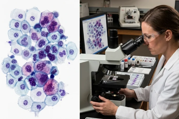
A tiny group of cells can reveal a surprising amount about what is happening inside the body. Changes in nuclear size, cell shape, chromatin, or the way cells cluster together may provide important clues about inflammation, infection, precancerous change, or malignancy.
This microscopic approach is called cytology, or cytopathology.
Unlike traditional histology, which examines cells within larger pieces of tissue, cytology focuses mainly on individual cells and small groups of cells obtained from body surfaces, fluids, or lesions.
Cytology can be used for both screening and diagnosis, with familiar examples including cervical cytology and fine-needle aspiration.
Understanding how cytology examines individual cells for signs of disease is especially useful for medical learners because it connects cellular morphology with clinical diagnosis.
The basic process is easy to follow:
Collect cells → prepare the specimen → stain the cells → examine morphology → identify abnormalities → perform additional tests when needed → report the findings
A few cells under a microscope may sometimes provide the first important clue that a disease is developing.
What Is Cytology?
Cytology is the microscopic examination of cells to identify abnormalities associated with disease.
The cells may come from epithelial surfaces, body fluids, organs, lymph nodes, or suspicious masses. Pathologists and cytology professionals evaluate characteristics such as cell size, shape, nuclear appearance, cytoplasmic features, and how cells are arranged relative to one another.
Cytology is especially well known for cancer screening and diagnosis, but its usefulness extends beyond malignancy.
Cytologic specimens can also contain inflammatory cells, infectious organisms, reactive changes, and other abnormalities. The Pap test, for example, can identify precancerous cervical changes and may also reveal inflammation or certain infections.
The central question is simple:
Do these cells look and behave like the normal cells expected at this site?
If they do not, the pattern of abnormalities helps determine what should happen next.
How Cells Are Collected for Cytology
Cytology does not rely on one collection method. The technique depends on where the suspected disease is located.
1. Exfoliative and Brush Cytology
Some cells naturally shed from epithelial surfaces or can be gently collected by brushing or scraping.
The Pap test is the best-known example. Cells are removed from the cervix and examined for abnormal changes that may represent precancer or cancer.
NCI describes cervical cytology as the collection of cervical cells so they can be checked for changes associated with cervical cancer development.
Similar principles can be applied to cells collected from other epithelial surfaces.
2. Body Fluid Cytology
Cells suspended in fluids can also be examined.
Urine, pleural fluid, peritoneal fluid, cerebrospinal fluid, and respiratory samples may contain cells that reveal inflammation, infection, or malignancy.
For example, cells in pleural fluid can be evaluated for malignant features.
CAP educational material highlights features such as a high nuclear-to-cytoplasmic ratio, irregular nuclear contours, and prominent nucleoli as findings that can raise concern for malignancy in the appropriate setting.
3. Fine-Needle Aspiration
Fine-needle aspiration, or FNA, uses a thin needle to remove cells and fluid from a lump or internal lesion.
It can be used for abnormalities involving areas such as the thyroid, lymph nodes, breast, pancreas, or lung.
Because the needle is small, FNA is generally less invasive than obtaining a larger core or surgical tissue specimen, while still providing material that may be useful for morphology and additional testing.
How Cytology Samples Become Microscope Slides
Collecting cells is only the first step.
The cells must then be preserved and prepared in a way that allows their microscopic features to remain recognizable.
Depending on the specimen, material may be spread directly onto glass slides, concentrated from a body fluid, processed using liquid-based methods, or prepared as a cell block.
The preparation is then stained.
One famous staining method is the Papanicolaou stain, commonly called the Pap stain. It provides excellent nuclear detail, which is particularly important because changes in the nucleus are among the most useful clues when evaluating abnormal cells.
Other staining methods may be used depending on the specimen and suspected disease.
Good specimen preparation matters because poor fixation, too few cells, excessive blood, crushing, or other artifacts can make interpretation difficult.
A cytology report may therefore describe a sample as inadequate or unsatisfactory if there is not enough reliable cellular material for interpretation.
NCI notes that cervical cytology results, for example, may be reported as unsatisfactory when the collected sample cannot be evaluated properly.
What Does a Cytopathologist Look for?
Under the microscope, cytology becomes a form of visual detective work.
The examiner does not simply ask whether a cell looks “strange.” Several features are considered together.
1. Nuclear Changes
The nucleus often provides some of the strongest clues.
Malignant or precancerous cells may show enlarged nuclei, irregular nuclear membranes, abnormal chromatin patterns, or prominent nucleoli.
A high nuclear-to-cytoplasmic ratio can also be concerning because some malignant cells contain relatively large nuclei compared with the amount of cytoplasm surrounding them.
2. Cell Size and Shape
Normal cells from a particular tissue usually fall within an expected range of shapes and sizes.
Marked variation between cells is known as pleomorphism. Significant pleomorphism may support a malignant diagnosis when combined with other abnormal features.
However, not every oddly shaped cell is cancerous. Infection, tissue repair, radiation, and inflammation can also create reactive cellular changes.
3. Cell Arrangement
Cytology also evaluates how cells relate to one another.
Some tumors produce cohesive clusters, while others release individual malignant cells. Gland-forming cancers may create recognizable three-dimensional groups, while lymphoid malignancies can produce more dispersed cell populations.
The surrounding background-including inflammation, mucus, necrotic material, or microorganisms-can provide additional clues.
Cytology Can Detect Precancer Before Invasive Cancer Develops
One of cytology’s most important roles is finding disease before it becomes clinically obvious.
Cervical cytology demonstrates this particularly well.
During a Pap test, cervical cells are examined for abnormal changes that may represent precancerous lesions. Detecting and treating significant precancerous abnormalities can prevent some of them from progressing to invasive cervical cancer.
This is an important distinction.
Cytology does not only look for fully developed cancer cells. It can identify dysplastic or precancerous cellular changes that indicate abnormal maturation and growth.
That makes cytology useful for screening because it may identify disease during a stage when intervention can occur before invasive malignancy develops.
Modern cervical screening increasingly incorporates HPV testing as well, so cytology may be interpreted together with molecular evidence of high-risk HPV infection depending on the screening strategy.
How Cytology Helps Diagnose Tumors
Outside screening, cytology often helps investigate a mass that has already been discovered.
Imagine an enlarged lymph node.
An FNA can collect cells from the node, allowing the cytopathologist to determine whether the population appears reactive, inflammatory, metastatic, or suspicious for a lymphoid malignancy.
The same approach can be used when imaging discovers a lung, thyroid, pancreatic, or breast lesion.
FNA specimens are especially valuable because they can provide diagnostic material while avoiding a larger procedure in suitable situations.
Modern cytology samples may also supply enough cellular material for ancillary testing, including molecular studies that help classify tumors and guide management.
Cytology can therefore move the investigation from:
“There is a mass”
to:
“These are the cells making up that mass.”
That is a major diagnostic step.
Cytology vs Histology: What Is the Difference?
Students often confuse cytology and histology, but the distinction is straightforward.
Cytology emphasizes individual cells and small cell groups.
Histology examines cells while preserving more of the surrounding tissue architecture.
Imagine looking at a city.
Cytology is like examining individual people: their appearance, characteristics, and small groups.
Histology is like viewing the neighborhood map as well. You can see where buildings, roads, and people are positioned relative to one another.
This architectural information can be essential.
For example, cytology may strongly suggest malignancy, but a tissue biopsy can show whether abnormal cells have invaded through normal anatomical boundaries.
This is one reason some positive cytology findings are followed by tissue biopsy before definitive management. NCI’s SEER training materials note that cytology can be used for screening or diagnosis, but biopsy is often performed to confirm cancer when appropriate.
The two techniques therefore complement rather than replace each other.
Additional Tests Can Extend What Cytology Tells Us
Modern cytology is not limited to visual examination.
When enough cells are available, immunocytochemistry, flow cytometry, molecular testing, or other ancillary studies may help clarify the diagnosis.
For example, antibodies can detect proteins that indicate whether malignant cells are epithelial, lymphoid, neuroendocrine, or another lineage.
Molecular testing can identify mutations or rearrangements associated with particular tumors. In lung cancer, small biopsy and cytology specimens can be strategically handled so they remain useful for predictive and prognostic ancillary testing.
CAP specifically emphasizes careful collection and triage of these specimens because modern therapy may depend on molecular information obtained from limited material.
This has transformed cytology.
A sample containing only a limited number of cells may now provide information about both diagnosis and tumor biology.
What Are the Limitations of Cytology?
Cytology is powerful, but it has limitations.
The biggest challenge is often sampling. The cells examined represent only the material that was successfully collected. If the needle or brush misses the abnormal area, the specimen may not reflect the true disease.
Small samples can also provide limited architectural information.
A pathologist may recognize malignant cells but still need a larger tissue biopsy to determine the exact tumor subtype, depth of invasion, or relationship to surrounding structures.
Reactive cells can create another challenge because inflammation and repair may produce changes that resemble malignancy.
For these reasons, cytology findings are interpreted alongside clinical history, imaging, laboratory data, and sometimes subsequent histologic biopsy.
A cytology result is therefore not simply a microscope opinion. It is one piece of a larger diagnostic puzzle.
A Simple Way to Remember Cytology
For medical learners, the workflow can be remembered as:
Collect → Prepare → Stain → Look → Compare → Classify → Confirm
Cells are first collected from a surface, fluid, or lesion.
They are prepared and stained so microscopic details become visible. The examiner then looks at nuclear features, cell shape, arrangement, and the surrounding background.
Those findings are compared with normal and known disease patterns.
The cells are then classified as normal, reactive, atypical, precancerous, suspicious, malignant, or another diagnosis depending on the specimen and reporting system.
When necessary, additional tests or tissue biopsy confirm and refine the interpretation.
Once you understand that sequence, cytology becomes much more than simply “looking at cells.”
Cytology examines individual cells for signs of disease by studying their microscopic appearance, including nuclear features, cell size and shape, arrangement, and surrounding background.
Cells may be collected through brushing, scraping, body fluids, or fine-needle aspiration.
Cytology can detect inflammation and infection, identify precancerous changes, investigate suspicious masses, and help diagnose malignancy. Modern specimens may also support immunologic or molecular testing.
Its main limitation is that a small collection of cells cannot always reveal the complete tissue architecture, so histologic biopsy may still be required.
For medical learners, remember the core question whenever you view a cytology slide: How do these cells differ from what should normally be here?
Follow their nuclei, cytoplasm, shape, arrangement, and background, and those tiny cells can tell a remarkably detailed story about disease.