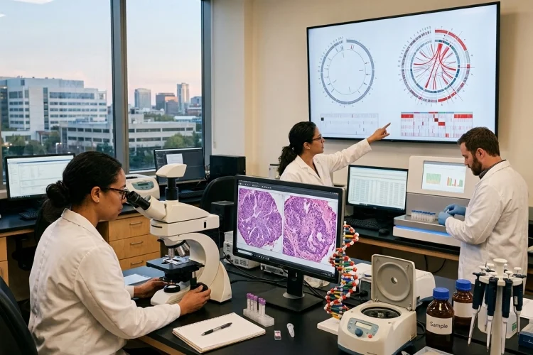
Two tumors can look almost identical under a microscope yet behave very differently. One may grow slowly, while another spreads rapidly. One might respond dramatically to a targeted drug, while the other ignores the same treatment.
The explanation may be hidden inside their DNA and RNA.
Molecular pathology studies genetic and molecular changes within diseased cells and combines those findings with traditional pathology.
In cancer, this approach can reveal mutations, gene fusions, amplifications, and other alterations that help explain why malignant cells grow, survive, or resist treatment.
Cancer itself is fundamentally a genetic disease caused by changes in genes involved in cellular growth and division.
Understanding how molecular pathology reveals cancer’s genetic drivers is becoming essential for medical learners because modern cancer diagnosis is no longer based on morphology alone.
The basic process can be viewed as:
Tumor sample → DNA or RNA analysis → genetic alteration → biological pathway → diagnosis or treatment implication
In other words, the microscope tells us what a tumor looks like. Molecular pathology helps explain what is driving it underneath the surface.
What Is Molecular Pathology in Cancer?
Molecular pathology examines disease at the level of DNA, RNA, genes, proteins, and molecular pathways.
In cancer care, the process usually begins with a tissue specimen obtained through biopsy or surgery. A pathologist first confirms that malignant cells are present and determines whether enough representative tumor is available for molecular analysis.
Laboratory testing can then search for specific genetic abnormalities or analyze many genes at once.
This information can complement histology by helping classify difficult tumors, identify recurrent genomic changes, and reveal potentially actionable abnormalities.
The College of American Pathologists notes that molecular features can sometimes help suggest a tumor’s type or origin and can influence how actionable variants are interpreted.
Molecular pathology therefore does not replace traditional pathology.
It adds another layer.
Morphology tells us what the cancer resembles; molecular testing tells us which biological instructions have changed.
What Are Cancer Genetic Drivers?
Cancer cells often contain many genetic alterations, but not every abnormality contributes equally to disease.
A driver alteration provides a growth or survival advantage that helps the malignant clone develop or expand. Other changes may simply accumulate as the tumor evolves without meaningfully driving its behavior.
Cancer-driving changes frequently involve genes controlling proliferation, cell death, DNA repair, or growth signaling. NCI identifies proto-oncogenes, tumor suppressor genes, and DNA repair genes as major groups affected during cancer development.
1. Oncogenes Push Growth Forward
Proto-oncogenes normally help regulate cell growth.
When mutations, amplification, or other changes make these genes excessively active, they can become oncogenes. The result may be continuous signaling that tells a cancer cell to grow or survive even when normal cells would stop.
2. Tumor Suppressor Genes Lose Their Brakes
Tumor suppressor genes normally restrain inappropriate proliferation or respond to cellular damage.
When their function is lost, an important protective brake disappears. If growth-promoting pathways are activated at the same time, the cell gains an even greater selective advantage.
Molecular pathology attempts to identify these alterations and determine whether they explain part of the tumor’s biology.
Molecular Pathology Detects More Than Simple Mutations
When people hear “genetic testing,” they often imagine searching for one misspelled DNA letter.
Cancer genomes are much more complicated.
A tumor may contain a single-nucleotide variant, where one DNA base has changed. Small insertions or deletions can also alter gene function.
Other cancers contain copy-number changes, where a gene has been amplified and exists in many extra copies. Structural alterations may break chromosomes and join pieces together, creating abnormal gene fusions.
For example, gene rearrangements involving ALK can act as important biomarkers in selected cancers, while other tumors are driven by abnormalities involving genes such as EGFR, BRAF, RET, or many others.
NCI’s tumor-marker resources list multiple molecular abnormalities that can influence cancer classification or treatment selection.
Molecular laboratories therefore need several techniques because one testing method cannot detect every type of genetic change equally well.
PCR and FISH Answer Focused Molecular Questions
Some molecular tests are designed to look for a specific abnormality rather than examine hundreds of genes.
1. PCR Detects Selected DNA or RNA Targets
Polymerase chain reaction, or PCR, can amplify specific genetic sequences so very small amounts of DNA or RNA become detectable.
This makes PCR useful when the laboratory already knows which molecular alteration it needs to investigate.
A targeted PCR assay can be fast and sensitive, but its major limitation is obvious: it usually finds only what it was designed to search for.
If a tumor contains an unexpected driver outside that target, the test may miss it.
2. FISH Visualizes Chromosomal Changes
Fluorescence in situ hybridization, or FISH, uses fluorescent probes that bind selected DNA regions inside cells.
It can reveal abnormalities such as gene amplification, deletion, or rearrangement.
FISH remains particularly useful in cancers where characteristic structural alterations have diagnostic or therapeutic significance. CAP notes that molecular methods including FISH and sequencing approaches have important roles in classifying tumors with recurrent genomic abnormalities.
These focused methods remain valuable even in the era of large sequencing panels because sometimes the clinical question is very specific.
Next-Generation Sequencing Looks at Many Genes at Once
Next-generation sequencing, commonly shortened to NGS, transformed molecular oncology by making it possible to study many genomic regions simultaneously.
Instead of performing separate tests for one mutation after another, an NGS panel may evaluate dozens or hundreds of cancer-associated genes in a single workflow.
Depending on the assay, NGS can detect combinations of single-nucleotide variants, small insertions and deletions, copy-number abnormalities, and structural changes. RNA-based approaches can also be particularly useful for identifying gene fusions.
CAP describes NGS as a technology capable of uncovering mutations and genomic variation that can improve understanding of disease biology and reveal opportunities for targeted treatment.
This becomes especially useful when several possible drivers could explain the cancer.
Imagine a lung adenocarcinoma.
Rather than testing individual genes one by one, a comprehensive panel may search simultaneously for multiple actionable alterations. The molecular report can then be integrated with histology and clinical information.
The result is something closer to a molecular map of the tumor.
Genetic Drivers Can Help Guide Targeted Therapy
Finding a cancer-driving alteration becomes especially valuable when a treatment exists that interferes with the abnormal pathway.
This is the principle behind targeted therapy.
Unlike traditional cytotoxic chemotherapy, which generally attacks rapidly dividing cells, targeted treatments are designed around molecular features that cancer cells depend on. NCI describes targeted therapy as treatment directed against changes in cancer cells that help them grow, divide, and spread.
Suppose molecular testing identifies an actionable mutation activating a particular signaling pathway.
A drug designed to inhibit that pathway may substantially reduce tumor growth in appropriately selected patients.
This produces the basic precision-oncology model:
Tumor → molecular test → actionable driver → matching therapy
Not every mutation has an available drug, and detecting a molecular alteration does not guarantee treatment success. But when a validated biomarker is strongly linked to response, molecular pathology can directly influence clinical decision-making.
Companion Diagnostics Connect Molecular Tests With Specific Treatments
Some molecular tests have such an important relationship with a therapy that they are classified as companion diagnostics.
The FDA defines a companion diagnostic as a medical device that provides information essential for the safe and effective use of a corresponding therapeutic product.
This means biomarker testing may be required before certain treatments are selected.
FDA’s current companion-diagnostic resources include oncology tests designed to identify molecular alterations associated with specific drugs or groups of drugs.
This relationship highlights how pathology has changed.
Historically, a pathology report might have primarily identified the tumor type and grade.
Today, the same diagnostic process may also answer:
Does this tumor contain the molecular target required for a particular therapy?
That is one of the clearest examples of molecular pathology influencing patient care.
Molecular Findings Can Help Classify Difficult Tumors
Molecular testing is not only about treatment.
Sometimes it helps determine what the tumor actually is.
Certain cancers contain highly characteristic gene fusions, amplifications, or mutational patterns. Detecting one of these abnormalities can support or refine a diagnosis when microscopic features overlap with several possible tumor types.
This is especially useful in some sarcomas, hematologic malignancies, brain tumors, and other molecularly defined neoplasms.
CAP has highlighted the importance of recurrent genomic alterations and mutational signatures in resolving complex cancer diagnoses, including situations where molecular findings may help indicate tumor origin or refine classification.
Modern pathology therefore increasingly follows an integrated model:
Histology + immunohistochemistry + molecular findings = final diagnosis
Rather than competing with microscopy, molecular testing makes the microscopic diagnosis more precise.
Tumors Can Develop Resistance to Targeted Treatment
Cancer does not remain biologically static.
A targeted therapy may initially eliminate cells carrying a vulnerable molecular pathway. However, other tumor cells may survive or acquire additional genetic changes that allow them to bypass the treatment.
This produces acquired resistance.
For example, a cancer may develop a new mutation that prevents a drug from binding effectively. Another clone may activate an alternative growth pathway and reduce its dependence on the original driver.
Molecular testing can sometimes identify these resistance mechanisms and reveal how a tumor has evolved during therapy.
This introduces an important concept for medical learners:
The molecular profile at diagnosis may not always be identical to the molecular profile after treatment.
Cancer is an evolving population of cells, and therapy itself creates selective pressure.
That is one reason repeat molecular testing can be clinically useful in selected settings.
Liquid Biopsy Can Reveal Tumor DNA Without a New Tissue Sample
Most molecular pathology begins with tissue, but tumors can also release genetic material into the bloodstream.
Fragments of tumor-derived DNA circulating in plasma are known as circulating tumor DNA, or ctDNA. Testing this material is one form of liquid biopsy.
Liquid biopsy may sometimes reveal cancer-associated molecular alterations without requiring another invasive tissue procedure. NCI has reported that blood-based genomic testing can identify tumor biomarkers that may help inform treatment options.
This approach is attractive for monitoring because blood samples can often be collected more easily than repeated tissue biopsies.
However, liquid biopsy has limitations. Some tumors release very little detectable DNA, and a negative plasma test does not always mean that an alteration is absent from the cancer.
Tissue and blood testing should therefore be viewed as complementary tools rather than automatic replacements for one another.
Molecular Results Need Careful Interpretation
Finding a variant is not the same as proving that it drives the cancer.
Large NGS panels can identify many genetic changes. Some are clearly pathogenic and clinically actionable. Others have uncertain biological significance.
The molecular pathologist therefore needs to interpret each finding in context.
Questions include:
Is this alteration known to drive this cancer?
Is it associated with an approved therapy?
Does its significance depend on the tumor type?
Could the alteration be inherited rather than restricted to the tumor?
Is there enough high-quality tumor material for the result to be reliable?
NCI emphasizes that each person’s cancer can carry a distinct biomarker pattern and that biomarker testing has important limitations; not every detected change leads to an effective treatment.
The final molecular report is therefore an interpretation of biological evidence-not simply a list of mutations.
A Simple Way to Understand Molecular Pathology
For medical learners, the process can be remembered as:
Sample → Extract → Detect → Interpret → Match → Monitor
First, representative tumor tissue is sampled.
DNA or RNA is then extracted and tested using methods such as PCR, FISH, or NGS.
The laboratory detects genetic alterations and determines which ones are likely to matter.
Pathologists then interpret those findings alongside morphology and clinical information.
If an actionable driver exists, the result may be matched with a targeted treatment or companion diagnostic strategy.
Finally, repeat tissue or liquid-biopsy testing may sometimes help monitor how the cancer changes over time.
Once this sequence is clear, molecular pathology becomes less about memorizing gene names and more about understanding how genetic information is converted into clinical decisions.
Molecular pathology reveals cancer’s genetic drivers by examining the DNA, RNA, and other molecular features hidden inside malignant cells.
PCR and FISH can answer focused genetic questions, while NGS can simultaneously explore many cancer-related genes.
These techniques identify mutations, amplifications, rearrangements, and gene fusions that may refine diagnosis, explain tumor biology, or reveal actionable targets for treatment.
The most important lesson for medical learners is that finding a mutation is only the beginning. The real question is what that alteration does.
When studying molecular oncology, follow the sequence: find the genetic change, identify the pathway it affects, decide whether it drives the cancer, and determine whether that vulnerability can be targeted.
That is where molecular pathology turns genetic information into practical cancer medicine.