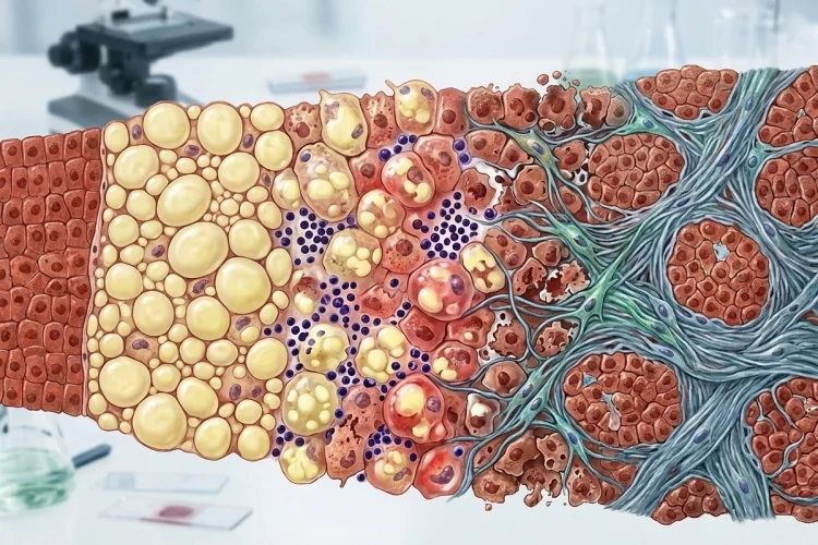
A fatty liver can sound fairly harmless. After all, the liver naturally handles fats every day. The problem begins when more fat enters or is produced in liver cells than the organ can safely process, store, or export.
This condition is now commonly called metabolic dysfunction-associated steatotic liver disease (MASLD) when liver fat occurs alongside cardiometabolic risk factors.
MASLD replaces the older term NAFLD, while metabolic dysfunction-associated steatohepatitis (MASH) replaces NASH.
Not everyone with fatty liver develops serious liver damage. In many people, fat accumulation causes little inflammation.
In others, however, cellular stress and immune activation develop, hepatocytes become injured, and the liver begins laying down scar tissue called fibrosis. Over years, advanced fibrosis may progress to cirrhosis.
Understanding how fatty liver progresses to inflammation and scarring is easier when you follow the process step by step: fat accumulation → cellular stress → inflammation → liver cell injury → fibrosis → possible cirrhosis.
What Happens During Fatty Liver?
The earliest change is hepatic steatosis, meaning excess fat accumulates inside hepatocytes, the main functional cells of the liver.
Under the microscope, these cells may contain large lipid droplets. In classic macrovesicular steatosis, a large fat vacuole can push the nucleus toward the edge of the hepatocyte.
In metabolic fatty liver disease, this process is closely connected with conditions such as insulin resistance, obesity, abnormal blood lipids, and type 2 diabetes. MASLD is specifically defined by hepatic steatosis together with at least one cardiometabolic risk factor.
At this point, there may be considerable liver fat without major inflammation or cellular injury.
That distinction matters. Fat accumulation alone is not the same thing as steatohepatitis.
Why Fat Starts Building Up in Hepatocytes
The liver sits at the center of carbohydrate and lipid metabolism, so it constantly receives and processes fatty acids.
When metabolic balance is disturbed, more fatty acids may reach the liver from adipose tissue. The liver may also create additional fatty acids from excess carbohydrate through de novo lipogenesis.
Normally, some fat is burned for energy, packaged into lipoproteins for export, or temporarily stored as triglycerides. Problems arise when the supply becomes greater than the liver’s ability to manage it safely.
Insulin resistance plays an important role in this metabolic environment and commonly occurs alongside MASLD.
AASLD notes that the newer terminology reflects the strong relationship between hepatic steatosis and metabolic factors such as visceral obesity, insulin resistance, and dyslipidemia.
For learners, imagine the hepatocyte as a warehouse. Temporary storage is manageable, but when deliveries keep arriving faster than products can be processed or shipped out, the warehouse becomes overloaded.
Fat Alone Is Not the Whole Problem
One of the most important ideas in fatty liver pathology is that simply seeing triglyceride droplets does not fully explain liver injury.
Some people have extensive steatosis without significant inflammation, while others develop hepatocyte damage and steatohepatitis.
NIDDK notes that experts still do not completely understand why some people remain at the relatively uncomplicated fatty-liver stage while others develop MASH/NASH.
The damaging process involves more than simple fat storage.
Excess fatty acids and their metabolites can disturb normal cellular metabolism. Mitochondrial stress, oxidative stress, altered lipid signaling, and other forms of cellular dysfunction can make hepatocytes increasingly vulnerable.
AASLD’s pathology guidance emphasizes that the biological injury in steatohepatitis is not simply caused by stored triglycerides; fatty acids and their metabolites are important contributors.
Once hepatocytes become stressed enough, simple steatosis can shift toward steatohepatitis.
When Fatty Liver Becomes MASH
MASH, formerly called NASH, means that fat accumulation is now accompanied by liver inflammation and hepatocyte injury.
Three classic microscopic features help pathologists recognize steatohepatitis: steatosis, lobular inflammation, and hepatocellular ballooning.
1. Hepatocyte Ballooning Shows Cell Injury
Ballooning describes hepatocytes that become enlarged and swollen because their internal structure has been damaged.
These cells look different from hepatocytes that simply contain a large fat droplet. Their cytoplasm may appear pale or granular, reflecting disruption of normal cellular architecture.
Ballooning is important because it tells the pathologist that the disease has moved beyond fat storage into active cellular injury.
2. Inflammation Joins the Process
Damaged and stressed hepatocytes produce signals that attract and activate immune cells.
Resident liver macrophages, including Kupffer cells, interact with recruited immune cells and inflammatory signaling pathways. This response is meant to deal with injured tissue, but persistent activation can create additional hepatocyte damage.
The result is a cycle:
Metabolic stress → hepatocyte injury → inflammation → more cellular stress and injury
When that cycle continues, the liver begins repairing repeated damage with scar tissue.
How Liver Injury Activates Scar-Producing Cells
The key transition from inflammation to fibrosis involves specialized cells called hepatic stellate cells.
Under normal conditions, stellate cells remain relatively quiet within the liver. Chronic injury changes their behavior. Signals released during hepatocyte damage and inflammation activate them, causing them to behave more like scar-producing myofibroblasts.
These activated cells begin producing extracellular matrix proteins, especially collagen.
Initially, this response is protective. Collagen provides structural support where repeated cell injury has disrupted normal tissue.
But when injury continues, collagen production does not switch off.
Scar tissue accumulates faster than it can be removed, and liver fibrosis develops.
This is the same basic repair principle seen elsewhere in pathology: persistent injury produces persistent healing, and persistent healing can eventually distort normal tissue.
How Fibrosis Spreads Through the Liver
Fibrosis does not usually transform the entire liver at once.
Early MASH-related fibrosis often develops in characteristic regions around hepatocytes and small vascular channels. As disease progresses, scar tissue can extend into additional areas of the liver.
Pathologists commonly describe fibrosis in stages. AASLD’s pathology guidance outlines progression from early perisinusoidal fibrosis through more extensive periportal involvement and bridging fibrosis, eventually reaching stage 4, or cirrhosis.
Bridging fibrosis means bands of scar tissue begin connecting normally separate regions.
That matters because the liver depends on a carefully organized relationship between hepatocytes, bile ducts, and blood vessels. As scar bands expand, normal architecture becomes increasingly distorted.
So the progression can be visualized as:
Localized fibrosis → expanding scar tissue → bridging fibrosis → architectural distortion → cirrhosis
Cirrhosis Is the Advanced Stage of Scarring
Cirrhosis develops when chronic fibrosis becomes extensive enough to permanently reorganize the liver.
Instead of smooth, normally arranged liver tissue, hepatocytes become surrounded by bands of fibrosis and form regenerative nodules. This distorted architecture interferes with normal blood flow and liver function.
MASH can progress to cirrhosis, and cirrhosis can eventually contribute to liver failure and increase the risk of liver cancer.
However, progression is not inevitable.
Many people with metabolic fatty liver never reach advanced fibrosis. The speed of progression also varies considerably between individuals.
Cardiometabolic conditions matter. Obesity and type 2 diabetes, for example, are strongly associated with MASLD, and people with multiple metabolic risk factors are more likely to develop the more active form of the disease.
Why Fatty Liver Can Be Difficult to Detect
One reason MASLD can progress unnoticed is that early disease may cause few or no obvious symptoms.
A person can accumulate liver fat for years without realizing it.
Routine imaging such as ultrasound, CT, or MRI can detect liver fat, but ordinary imaging cannot reliably tell whether inflammation or fibrosis is present.
Tests such as FIB-4 can help identify people at risk of advanced fibrosis, while elastography can estimate liver stiffness associated with scarring.
In selected cases, doctors may use a liver biopsy.
A biopsy allows a pathologist to directly evaluate steatosis, hepatocyte ballooning, inflammation, and fibrosis. NIDDK notes that biopsy can establish steatohepatitis and clearly assess disease severity, although it is not required for everyone with suspected fatty liver disease.
This is why simply discovering “fat on the liver” does not tell the entire story. The clinically important question is often how much injury and fibrosis are present.
Can the Progression Be Slowed or Reversed?
Fatty liver disease is dynamic rather than a one-way pathway.
Reducing the metabolic stress driving the disease can decrease liver fat and may also improve inflammation and fibrosis. NIDDK notes that weight loss can reduce hepatic fat and can also improve inflammation and scarring associated with MASH/NASH.
The possibility of improvement is especially important in earlier stages, before severe architectural distortion has developed.
Modern management therefore looks beyond the liver alone. Cardiometabolic factors such as body weight, glucose control, blood pressure, and lipid abnormalities are important parts of evaluating people with MASLD.
Current liver guidelines likewise approach MASLD as a disease closely connected with overall metabolic health.
For medical learners, this reinforces a broader lesson: pathology is often reversible when the injurious stimulus can be reduced before permanent structural damage occurs.
A Simple Way to Remember Fatty Liver Progression
Instead of memorizing MASLD, MASH, and fibrosis as separate diagnoses, connect them into one biological story:
Metabolic dysfunction → excess liver fat → lipotoxic stress → hepatocyte ballooning and injury → inflammation → stellate cell activation → collagen deposition → fibrosis → cirrhosis
Not every patient travels through every stage.
Some remain with relatively uncomplicated steatosis for years, while others develop inflammation and progressive fibrosis.
The key turning point is cellular injury.
Once hepatocytes are being damaged rather than simply storing fat, inflammatory and wound-healing pathways become activated. If the source of injury continues, the healing response itself begins reshaping the liver.
Fatty liver progresses to inflammation and scarring when excess lipid accumulation becomes part of a wider cycle of metabolic stress, hepatocyte injury, immune activation, and abnormal tissue repair.
Simple steatosis may cause little liver damage, but MASH includes fat, inflammation, and ballooning injury. Persistent damage activates scar-producing cells, leading to collagen deposition, fibrosis, bridging scars, and potentially cirrhosis.
For medical learners, the most useful sequence is straightforward: fat accumulates, cells become stressed, inflammation develops, and repeated repair creates scar tissue.
When studying fatty liver disease, do not stop at asking how much fat is present. Ask whether hepatocytes are being injured and whether fibrosis has begun-because those changes reveal much more about where the disease is heading.