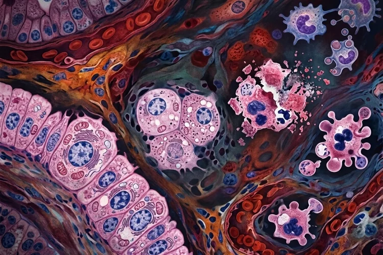
Cells may be tiny, but they are constantly making adjustments to survive. Every day, they deal with changes in oxygen supply, nutrients, hormones, workload, temperature, toxins, infections, and many other environmental pressures.
So, how do cells respond to stress, injury, and changing conditions? The answer depends largely on how intense the stress is, how long it lasts, and what type of cell is affected.
A mild challenge may cause the cell to adapt. A stronger disturbance can produce reversible cell injury, while severe or prolonged damage may cross the point of no return and cause cell death.
This progression is one of the most important foundations of general pathology. Once you understand it, concepts such as hypertrophy, hyperplasia, atrophy, metaplasia, hypoxic injury, necrosis, and apoptosis become much easier to connect.
Instead of memorizing each process separately, think of them as different answers to the same question: What can a cell do when its normal environment changes?
Why Cells Need to Adapt
A healthy cell normally exists within a relatively stable range of conditions known as homeostasis. However, the environment around a cell rarely stays perfectly constant.
Hormone levels change. Nutrient availability changes. Tissues may suddenly have to work harder. Cells may also experience reduced oxygen, inflammation, infection, mechanical pressure, or chemical exposure.
When these challenges remain within manageable limits, cells can modify their structure or function to maintain survival. Cellular adaptation is generally reversible if the stimulus disappears.
A useful way to imagine this is to think of a cell as a small factory. If demand increases, the factory may expand production. If resources become limited, it may reduce its activity. If the surrounding environment becomes uncomfortable, it may even change the type of machinery it uses.
In pathology, several major adaptations explain these changes: hypertrophy, hyperplasia, atrophy, and metaplasia.
Hypertrophy: When Cells Become Larger
Hypertrophy means an increase in the size of individual cells, usually resulting in enlargement of the affected tissue or organ. It is especially important in tissues where mature cells have limited ability to divide.
Skeletal muscle provides an easy example. Repeated resistance exercise places greater mechanical demand on muscle fibers. In response, the cells increase their protein content and become larger.
Hypertrophy can therefore be physiological and useful.
However, it can also be pathological.
Consider chronic high blood pressure. The left ventricle of the heart must generate greater pressure to pump blood against increased resistance. Cardiac muscle cells may respond by becoming larger, producing left ventricular hypertrophy.
Initially, this adaptation helps the heart manage the increased workload. But if excessive pressure continues for years, the same adaptation can eventually contribute to impaired cardiac function.
This introduces an important pathology principle: an adaptation that helps initially may become harmful when the stress continues for too long.
Hyperplasia: When Cell Numbers Increase
While hypertrophy increases cell size, hyperplasia increases the number of cells within a tissue.
Hyperplasia can occur when cells are capable of dividing and receive signals that stimulate proliferation. Like hypertrophy, it can be either physiological or pathological.
A classic physiological example occurs during pregnancy. Hormonal stimulation contributes to both enlargement and an increase in the number of smooth muscle cells within the uterus. In other words, hypertrophy and hyperplasia can occur together.
Pathological hyperplasia occurs when excessive hormonal or growth signals produce too much cell proliferation. Benign prostatic hyperplasia, for example, involves nonmalignant proliferation of prostate tissue and becomes increasingly common with advancing age.
An important distinction for medical learners is that hyperplasia is controlled proliferation, unlike cancer, where abnormal cells develop autonomous and dysregulated growth characteristics.
Still, some forms of pathological hyperplasia can create conditions in which additional genetic or cellular abnormalities become clinically important.
Atrophy: When Cells Reduce Their Size
Sometimes survival requires doing less rather than more.
Atrophy refers primarily to a decrease in cell size, often accompanied in tissues by reduced functional capacity and, in some settings, loss of cells. It can happen when a tissue receives less stimulation, fewer nutrients, reduced blood flow, or decreased workload.
Muscle immobilization offers a familiar example. If someone wears a cast for several weeks after a fracture, the muscles inside the immobilized limb become smaller because they are being used less.
Atrophy may also occur with aging, loss of hormonal stimulation, inadequate nutrition, denervation, or chronic reduction in blood supply.
From the cell’s perspective, shrinking can be an energy-saving strategy. Maintaining a large amount of cellular machinery requires resources. When demand falls or nutrients become scarce, reducing cellular components can help preserve survival.
The key idea is that atrophy does not automatically mean the cell is dead. It represents an adjustment to a changed environment. If favorable conditions return soon enough, some forms of atrophy can improve.
Metaplasia: When One Cell Type Is Replaced by Another
Metaplasia is a particularly interesting adaptation because the tissue changes the type of mature cells it produces.
More precisely, one differentiated cell type is replaced by another differentiated cell type that is better able to tolerate a particular chronic stress.
One well-known example occurs in Barrett esophagus. The normal esophagus is lined primarily by stratified squamous epithelium.
With chronic gastroesophageal reflux, cells in the lower esophagus may be replaced by intestinal-type columnar epithelium containing characteristic goblet cells.
Why would this happen?
The new epithelium is better suited to dealing with the abnormal chemical environment created by repeated reflux. In that sense, metaplasia is protective.
But there is a catch.
Persistent pathological metaplasia can create a setting in which dysplasia and eventually malignancy become more likely. Barrett esophagus, for example, is associated with an increased risk of esophageal adenocarcinoma.
So again, adaptation may protect cells in the short term while producing new risks over time.
What Happens When Adaptation Is Not Enough?
Cells can only compensate up to a certain point.
If a stress becomes too severe, arrives too quickly, or continues for too long, cellular adaptation may no longer be enough. The cell then enters a state of cell injury.
Many things can cause injury, including reduced oxygen availability, ischemia, toxins, infections, immune reactions, nutritional imbalance, extreme temperatures, physical trauma, and genetic abnormalities.
One of the most important mechanisms is ATP depletion.
Cells require ATP to maintain ion pumps in their membranes. When oxygen delivery falls significantly, mitochondrial ATP production decreases. Energy-dependent pumps begin to fail, sodium accumulates inside the cell, and water follows it. The result can be cellular swelling.
Mitochondrial dysfunction, increased intracellular calcium, membrane damage, protein misfolding, DNA injury, and oxidative stress can also contribute to cellular damage.
These mechanisms often interact rather than occurring independently.
Reversible vs Irreversible Cell Injury
An essential distinction in pathology is whether an injured cell can still recover.
1. Reversible Cell Injury
During reversible cell injury, the cell has been damaged but has not yet crossed the point where recovery is impossible.
Cellular swelling is one of the classic early changes. ATP depletion interferes with membrane ion pumps, allowing sodium and water to enter the cell.
Other changes may affect mitochondria, the endoplasmic reticulum, ribosomes, and cellular metabolism.
If the harmful stimulus is removed early enough, the cell may restore its normal structure and function. This is why the duration and severity of an injury matter as much as the type of injury itself.
2. Irreversible Cell Injury
If damage continues, the cell eventually reaches a stage from which recovery is no longer possible.
Severe mitochondrial dysfunction and extensive damage to cellular and organelle membranes are important features associated with irreversible injury.
At that point, the cell will die.
A useful sequence for students is:
Stress → Adaptation → Reversible injury → Irreversible injury → Cell death
Not every disease follows this pathway perfectly, but it provides an excellent framework for understanding many pathological processes.
Apoptosis and Necrosis: Two Important Forms of Cell Death
When cells can no longer survive, the final outcome may involve several types of cell death. Two concepts medical learners encounter constantly are apoptosis and necrosis.
1. Apoptosis
Apoptosis is a tightly regulated form of programmed cell death. It plays normal roles in development and tissue maintenance while also helping eliminate cells that are damaged or potentially harmful.
During apoptosis, the cell essentially dismantles itself in an organized way. Chromatin condenses, cellular components are packaged into membrane-bound fragments, and these fragments can be removed by neighboring cells or phagocytes.
Because the membrane remains relatively controlled during this process, apoptosis typically generates much less surrounding inflammation than classic necrosis.
2. Necrosis
Necrosis commonly follows severe pathological injury.
Cells undergoing necrosis typically swell, lose membrane integrity, and eventually release intracellular material into surrounding tissue. That leakage can trigger an inflammatory response.
For example, when blood flow to part of the heart is interrupted long enough during myocardial infarction, severe ischemic injury can result in necrosis of cardiac muscle cells.
For learners, the easiest basic comparison is this:
Apoptosis is organized cellular removal, while necrosis involves destructive cellular injury with membrane breakdown and inflammation.
Modern research recognizes additional regulated cell-death pathways, including necroptosis, pyroptosis, and ferroptosis, so the biology is more complex than a simple apoptosis-versus-necrosis division.
How to Study Cellular Adaptation and Injury
The easiest way to learn this topic is not to memorize every definition independently.
Instead, ask a sequence of questions whenever you encounter a pathological condition.
First, what is the stressor? Is it increased workload, reduced blood supply, hormonal stimulation, infection, or a toxin?
Next, ask how the cell responds. Does it become larger, increase in number, shrink, or change phenotype?
Then determine whether the response is still adaptive or whether genuine injury has begun.
Finally, ask whether the injury is reversible and what happens if it continues.
For example:
Hypertension → Increased cardiac workload → Myocyte hypertrophy → Left ventricular hypertrophy → Prolonged stress → Possible cardiac dysfunction
This cause-and-effect approach turns pathology from a collection of vocabulary terms into a biological story.
Cells constantly adjust to changes in workload, nutrients, hormones, oxygen, and environmental stress. When the challenge is manageable, cellular adaptation through hypertrophy, hyperplasia, atrophy, or metaplasia can help maintain survival.
When stress exceeds the cell’s adaptive capacity, however, cell injury begins. Mild injury may remain reversible, while severe or prolonged damage can become irreversible and ultimately result in apoptosis, necrosis, or other forms of cell death.
For medical learners, the most useful habit is to follow the sequence rather than simply memorize definitions: identify the stress, understand the adaptation, recognize the injury, and predict the outcome.
The next time you study a disease, ask what happened at the cellular level first. That single question can make the entire pathophysiology much easier to understand.