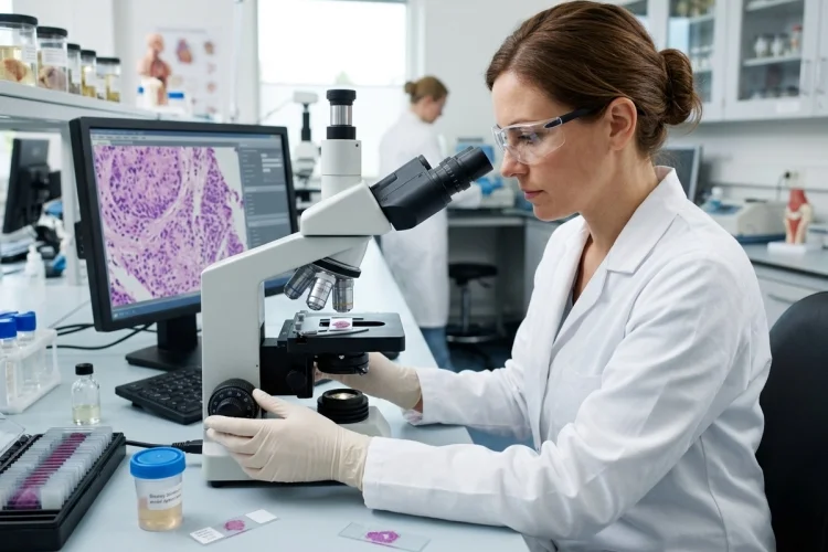
A scan can reveal a suspicious lump. A blood test can suggest that something is wrong. Symptoms can point doctors toward several possible diseases. But sometimes the clearest answers come from looking directly at the affected cells and tissues.
That is where a biopsy sample becomes incredibly valuable.
A biopsy involves removing cells or tissue from the body so they can be examined for signs of disease. A pathologist studies the sample under a microscope and may use additional laboratory techniques to determine what is happening inside the tissue.
Biopsies are especially important in cancer diagnosis, but they are also used to investigate infections, inflammatory disorders, skin diseases, organ abnormalities, and many other medical conditions.
Understanding how biopsy samples help pathologists diagnose human disease gives medical learners a useful look behind the scenes of diagnostic medicine.
The process is much more than simply placing tissue under a microscope. It involves careful sampling, preservation, processing, microscopic interpretation, and sometimes molecular testing before a final diagnosis can be made.
What Is a Biopsy Sample?
A biopsy sample is a small amount of cells or tissue collected from an area of the body that needs closer examination.
The reason for taking a biopsy varies. A doctor may want to investigate a suspicious mass, an abnormal area seen on imaging, an unusual skin lesion, persistent inflammation, or tissue changes that cannot be fully explained by other tests.
Pathologists examine these specimens for evidence of abnormal cellular structure, tissue damage, inflammation, infection, precancerous changes, or malignancy.
MedlinePlus notes that biopsies can be performed throughout the body and are used for conditions far beyond cancer alone.
A biopsy can therefore answer a very practical question:
What is this abnormal tissue actually made of?
That information can dramatically narrow the differential diagnosis.
Different Biopsy Methods Collect Different Samples
Not every biopsy is performed in the same way. The technique depends on the organ, location of the abnormality, size of the lesion, and the amount of tissue needed.
1. Needle Biopsy
A needle biopsy removes cells or tissue through a needle inserted into the suspicious area.
Fine-needle aspiration typically collects cells and fluid, while a core needle biopsy removes a small cylinder of tissue. Core samples preserve more of the tissue architecture, allowing pathologists to examine how cells are arranged in relation to one another.
2. Endoscopic Biopsy
Some internal organs can be sampled using an endoscope.
For example, a doctor performing an upper gastrointestinal endoscopy or colonoscopy can pass small instruments through the endoscope and remove tissue from an abnormal area. NCI notes that endoscopic techniques are among several common ways biopsy specimens can be obtained.
3. Incisional and Excisional Biopsy
An incisional biopsy removes only part of a lesion, while an excisional biopsy removes the entire lesion.
Larger surgical specimens may provide additional information that a small biopsy cannot, including the overall size of a tumor, its relationship to surrounding tissues, and whether surgical margins contain abnormal cells.
The key principle is simple: the quality of the diagnosis depends partly on obtaining an appropriate and representative sample.
What Happens to a Biopsy in the Pathology Laboratory?
Once the sample reaches the pathology laboratory, it has to be prepared carefully before a pathologist can study it.
For many tissue specimens, the first important step is fixation. Formalin is commonly used to preserve tissue structure and prevent cells from rapidly breaking down after removal.
The specimen may then undergo additional processing in which water is removed and replaced with paraffin wax. The tissue becomes embedded in a paraffin block that can be cut into extremely thin sections.
These sections are placed onto glass slides and stained so cellular structures become easier to recognize under a microscope.
One of the most familiar stains in pathology is hematoxylin and eosin, commonly shortened to H&E.
Hematoxylin generally makes nuclei appear blue to purple, while eosin highlights many proteins and cytoplasmic structures in shades of pink. This contrast allows pathologists to examine tissue architecture, cell shape, nuclear appearance, inflammation, necrosis, and many other features.
Johns Hopkins describes histologic sections as very thin, stained slices of specimens placed on glass slides for microscopic examination.
What Does a Pathologist Look for Under the Microscope?
This is where diagnostic pathology becomes something like biological detective work.
A pathologist rarely asks only whether cells look “normal” or “abnormal.” Instead, multiple features are considered together.
The pathologist examines the overall architecture of the tissue. Are cells arranged in their normal pattern? Have normal tissue boundaries disappeared? Are abnormal glands, nests, sheets, or other structures present?
Individual cell characteristics are also important.
The pathologist may evaluate cell size and shape, nuclear appearance, mitotic activity, differentiation, inflammation, tissue destruction, fibrosis, microorganisms, or abnormal deposits.
In tumors, these details may help determine whether a lesion is benign or malignant and what type of tumor is present.
NCI explains that pathology reports may identify features such as tumor type, grade, lymph-node involvement, margin status, and other findings relevant to diagnosis and treatment.
In other words, morphology provides the first major set of clues.
Special Stains and Immunohistochemistry Add More Clues
Sometimes the routine H&E slide gives a clear answer. Other times, several diseases look surprisingly similar under the microscope.
That is when additional tests become useful.
Special stains can highlight substances, microorganisms, connective tissue components, or other structures that may be difficult to identify with routine staining.
Another powerful technique is immunohistochemistry, often abbreviated as IHC.
IHC uses antibodies that bind to particular proteins inside or on the surface of cells. The resulting staining pattern can help determine what type of cells make up a lesion.
For example, two tumors may look similar under routine microscopy but express very different proteins. An IHC panel can help the pathologist determine whether a tumor is likely derived from epithelial cells, lymphocytes, melanocytes, muscle cells, or another cell lineage.
NCI notes that immunohistochemical and immunophenotyping markers are widely used to distinguish among different types of cancer.
This is why modern diagnosis often combines morphology with molecular identity.
Molecular Testing Can Reveal Changes Invisible Under the Microscope
Cells may look similar while carrying very different genetic abnormalities.
Modern pathology therefore increasingly uses molecular testing to analyze DNA, RNA, chromosomes, or specific proteins within biopsy tissue. NCI notes that molecular analysis of tissue can refine cancer classification and help guide treatment decisions.
Some mutations or gene rearrangements are strongly associated with particular tumor types. Others predict whether a patient may benefit from a targeted therapy.
For example, testing tumor tissue for certain biomarkers can provide information about treatment selection or prognosis. NCI lists numerous molecular tumor markers that are evaluated in blood, bone marrow, or tumor tissue for these purposes.
This means that a biopsy can provide much more information than simply “cancer” or “not cancer.”
In modern medicine, a single tissue sample may reveal the disease type, subtype, biological behavior, and potential therapeutic targets.
Biopsies Diagnose More Than Cancer
Cancer may be the condition most commonly associated with biopsies, but pathologists diagnose a much wider range of diseases.
A skin biopsy, for example, may reveal an inflammatory skin disorder, fungal infection, autoimmune process, or benign growth.
Biopsies of organs such as the liver, kidney, intestine, or lung can help identify patterns of inflammation, fibrosis, infection, immune-mediated damage, or abnormal protein accumulation.
The microscopic pattern matters because many diseases produce characteristic combinations of cellular injury and tissue response.
Imagine a liver biopsy.
Instead of simply seeing “damaged liver,” the pathologist may evaluate where inflammation is occurring, whether hepatocytes are injured, how much fibrosis is present, and whether unusual deposits or other abnormalities can be identified.
The result helps transform a broad clinical problem into a more specific pathological diagnosis.
The Pathology Report Turns Tissue Findings Into Clinical Information
After examining the sample and completing any necessary additional tests, the pathologist creates a pathology report.
A typical report may include information about the specimen, a gross description, microscopic findings, the diagnosis, and comments or recommendations for further testing.
Johns Hopkins describes the diagnosis section as the main conclusion based on what was found in the specimen.
For cancer specimens, the report may also include clinically important information such as tumor type, grade, margins, lymph-node status, and biomarker results. These findings can influence staging and treatment planning.
The pathologist therefore acts as a bridge between what the tissue looks like biologically and what the treating physician needs to know clinically.
A few millimeters of tissue can sometimes change an entire treatment plan.
Why Biopsy Results Are Not Always Simple
Although biopsy is an extremely powerful diagnostic tool, it also has limitations.
A biopsy only represents the tissue that was actually sampled. If a lesion is large or biologically heterogeneous, a small specimen may not contain every abnormal area.
Specimen quality matters too.
Poor preservation, crushing of tissue during collection, insufficient tissue, or sampling an area dominated by necrosis can make diagnosis more difficult. Some specimens also need to be reserved carefully for additional tests such as immunohistochemistry, cytogenetics, or molecular analysis.
CAP guidance emphasizes the importance of appropriate specimen handling and tissue allocation when ancillary studies may be required.
Pathologists therefore interpret a biopsy together with relevant clinical history, imaging findings, laboratory information, and the exact site from which the sample was obtained.
When the available tissue cannot answer the clinical question confidently, additional sampling may sometimes be necessary.
A Simple Way to Remember the Biopsy Process
For medical learners, the entire pathway can be remembered as:
Sample → Preserve → Process → Stain → Examine → Test → Diagnose
A clinician first obtains representative tissue. The laboratory preserves and prepares it. The pathologist evaluates its microscopic architecture and cellular features.
If morphology alone is not enough, special stains, immunohistochemistry, or molecular tests provide additional clues.
Finally, all the findings are integrated into a pathology report that helps the medical team understand what disease is present and, in many cases, how it should be managed.
This approach makes biopsy pathology much easier to understand than memorizing laboratory techniques individually.
Biopsy samples help pathologists diagnose human disease by allowing abnormal tissues to be examined directly at the cellular and microscopic level.
After a specimen is collected, it is carefully preserved, processed, sectioned, stained, and examined.
Routine morphology may provide the diagnosis, while immunohistochemistry, special stains, or molecular testing can reveal additional information that is impossible to see with the naked eye alone.
Biopsies can identify cancer, infection, inflammation, fibrosis, and many other pathological processes. For medical learners, the key is to follow the complete journey: from patient to specimen, from specimen to slide, and from slide to diagnosis.
The next time you read a pathology report, try tracing each conclusion back to the tissue evidence that made it possible.