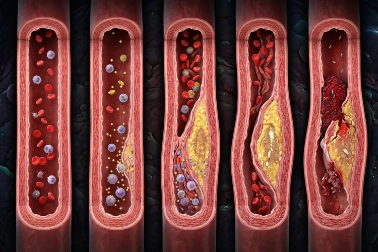
Atherosclerosis does not usually appear overnight. Long before someone develops chest pain, a heart attack, or a stroke, subtle changes may already be taking place inside the walls of their arteries.
At first, the artery may look almost normal. Over time, however, cholesterol-rich particles can accumulate within the arterial wall, inflammatory cells arrive, and a growing lesion begins to form.
The body attempts to contain the problem by building fibrous tissue around it, eventually creating what we recognize as an atherosclerotic plaque.
This process can slowly narrow an artery, but an even more dangerous event can occur when a plaque becomes unstable and triggers formation of a blood clot.
Understanding how atherosclerosis develops inside arteries over time is especially important for medical learners because it connects lipid metabolism, endothelial dysfunction, inflammation, thrombosis, and cardiovascular disease.
Rather than thinking of atherosclerosis as simply “fat blocking an artery,” it is more useful to see it as a long-term biological process involving lipids, immune cells, vascular cells, and tissue repair.
What Is Atherosclerosis?
Atherosclerosis is a disease in which cholesterol, fat, blood cells, calcium, and other substances gradually accumulate within artery walls and form plaque.
As plaques grow, affected arteries may become thicker, stiffer, and narrower, reducing the flow of oxygen-rich blood to tissues.
It is important to distinguish atherosclerosis from the broader term arteriosclerosis. Arteriosclerosis generally refers to hardening or loss of elasticity of arteries, while atherosclerosis specifically involves plaque formation within the arterial wall.
Atherosclerosis can affect arteries throughout the body. When coronary arteries are involved, it can contribute to coronary heart disease.
Plaques affecting arteries supplying the brain can contribute to ischemic stroke, while disease in arteries of the limbs can lead to peripheral artery disease.
The disease develops slowly, often over many years.
The Process Begins at the Endothelium
The inner surface of an artery is lined by a thin layer of cells called the endothelium.
This lining is not simply a passive covering. Endothelial cells help regulate vascular tone, blood clotting, inflammation, and movement of substances between blood and tissues.
In atherosclerosis-prone areas, changes in normal endothelial function can create an environment that encourages lipid retention and inflammatory cell recruitment.
Endothelial dysfunction is therefore considered an important early contributor to the development and progression of atherosclerotic lesions.
Several cardiovascular risk factors can promote this unfavorable environment. These include elevated cholesterol, high blood pressure, smoking, diabetes, aging, and other metabolic or genetic factors.
Having multiple risk factors further increases the likelihood of atherosclerosis and its complications.
For learners, an easy starting point is:
Risk factors → endothelial dysfunction → easier lipid accumulation and inflammation
That sets the stage for plaque formation.
LDL Particles Begin Accumulating in the Arterial Wall
Low-density lipoprotein, or LDL, carries cholesterol through the bloodstream.
LDL itself has normal biological functions, but persistently elevated concentrations increase the chance that cholesterol-containing particles will become retained within susceptible areas of the arterial wall.
Elevated LDL cholesterol is strongly associated with the development of atherosclerotic cardiovascular disease.
Once retained within the vessel wall, lipoproteins can undergo chemical modifications and interact with surrounding cells.
The important idea is that cholesterol is no longer simply circulating through the artery. It has become trapped inside the arterial intima, where it can stimulate a local inflammatory response.
The body recognizes that something abnormal is occurring and begins recruiting immune cells.
This transforms what began as lipid accumulation into something much more complicated: chronic inflammation within the artery wall.
Macrophages Turn Into Foam Cells
One of the classic cellular events in atherosclerosis involves monocytes and macrophages.
Circulating monocytes are recruited into the affected arterial wall, where they can differentiate into macrophages. These macrophages take up large quantities of cholesterol-rich lipoproteins.
As lipids accumulate inside them, the macrophages develop a characteristic foamy appearance under the microscope. They are therefore called foam cells. NHLBI describes cholesterol-laden macrophages as an important component of atherosclerotic plaques.
Collections of lipid-containing foam cells help produce an early lesion known as a fatty streak.
A fatty streak does not necessarily cause significant obstruction. At this stage, blood may still move through the artery without noticeable difficulty.
However, continuing lipid retention and inflammation can cause the lesion to grow.
This is one reason atherosclerosis can remain clinically silent for such a long time. Biological changes may already be occurring even when the person has no symptoms.
A Fatty Streak Becomes a Fibrous Plaque
As atherosclerosis progresses, it becomes more than a collection of foam cells.
Vascular smooth muscle cells participate in the response. They can migrate toward the inner part of the artery, proliferate, and produce extracellular matrix components such as collagen.
Over time, this creates a fibrous cap over a deeper lipid-rich region.
The plaque may eventually contain cholesterol, inflammatory cells, connective tissue, smooth muscle cells, cellular debris, and calcium. As it becomes larger, the artery wall thickens and the space available for blood flow may decrease.
A simplified mature plaque can therefore be imagined as having two major regions:
Fibrous cap → protective connective tissue layer
Lipid or necrotic core → cholesterol, debris, and dead cells beneath it
The fibrous cap is particularly important because it separates highly thrombogenic material inside the plaque from circulating blood.
In a sense, the body is trying to contain a chronic inflammatory problem by building a wall around it.
Plaques Can Grow Without Causing Symptoms
One surprising feature of atherosclerosis is that plaque does not always produce symptoms immediately.
Arteries can compensate for gradual changes for a long time, and symptoms typically depend on the artery involved and how much blood flow has been affected.
NHLBI notes that many people do not know they have atherosclerosis until plaque buildup causes significant narrowing or complications.
If coronary arteries become significantly narrowed, blood supply may become inadequate when the heart needs more oxygen, such as during physical activity.
If atherosclerosis affects the arteries supplying the legs, reduced blood flow can contribute to symptoms of peripheral artery disease.
However, the amount of narrowing is only part of the story.
A plaque that produces relatively modest narrowing may still become dangerous if its surface suddenly becomes disrupted.
That is why plaque stability matters just as much as plaque size.
Why Some Atherosclerotic Plaques Become Dangerous
Not every plaque has the same structure or biological behavior.
Some plaques develop relatively thick fibrous caps and may remain stable for long periods. Others contain large lipid-rich cores, substantial inflammatory activity, and thinner or mechanically weaker caps.
Ongoing inflammation can affect smooth muscle cells and extracellular matrix within the plaque, potentially reducing the strength of the fibrous cap. Lipid accumulation also contributes to inflammatory processes associated with plaque vulnerability.
If the protective surface becomes disrupted, the situation changes rapidly.
Material that had been hidden within the plaque becomes exposed to circulating blood. Platelets and coagulation pathways can then be activated.
A process that developed silently over decades can suddenly become an emergency within minutes.
Plaque Rupture Can Trigger a Blood Clot
One of the most dangerous complications of atherosclerosis is thrombosis.
When a vulnerable plaque ruptures-or when its surface is disrupted through related mechanisms-the blood encounters material that promotes clot formation. Platelets accumulate, coagulation becomes activated, and a thrombus can develop over the plaque.
If that clot becomes large enough, it may suddenly obstruct blood flow.
In a coronary artery, acute interruption of blood supply can produce a myocardial infarction. In an artery supplying part of the brain, blockage may lead to an ischemic stroke. Atherosclerotic plaque buildup also increases the likelihood of blood clots forming within affected arteries.
This explains an important clinical concept:
Atherosclerosis is chronic, but its complications can be sudden.
The plaque may have taken years to develop, yet thrombosis can transform a previously stable condition into a life-threatening event very quickly.
Why Atherosclerosis Is More Than a Cholesterol Storage Problem
It is tempting to imagine an atherosclerotic artery as a pipe gradually filling with grease.
That analogy is useful only up to a point.
Atherosclerosis is biologically active. The plaque contains living vascular cells, immune cells, extracellular matrix, cholesterol-rich material, and signaling molecules. Inflammation influences both plaque progression and its potential instability.
The arterial wall itself also changes.
Smooth muscle cells respond, extracellular matrix is remodeled, cells die, lipid accumulates, and some advanced plaques can become calcified.
A better model is therefore:
Lipid retention → inflammation → foam cells → tissue remodeling → fibrous plaque → possible plaque instability → thrombosis
This sequence brings together many subjects that medical learners encounter separately in pathology, immunology, and cardiovascular physiology.
What Makes Atherosclerosis Progress Faster?
The speed and severity of atherosclerosis vary significantly between individuals.
High LDL cholesterol is a major risk factor. High blood pressure, tobacco exposure, diabetes, obesity, physical inactivity, increasing age, family history, and other factors may also contribute to cardiovascular risk.
These factors do not necessarily work independently.
For example, someone with high LDL cholesterol plus hypertension and smoking exposure may have greater overall risk than someone with only one risk factor.
This also explains why atherosclerosis prevention focuses heavily on managing modifiable cardiovascular risks.
Depending on an individual’s risk, this may involve dietary changes, physical activity, smoking cessation, blood pressure management, diabetes management, and cholesterol-lowering therapy under medical guidance.
From a pathology perspective, reducing these risk factors means reducing the biological forces that encourage plaque formation and progression.
A Simple Way to Remember Atherosclerosis Development
For medical learners, the entire process can be reduced to a useful chain:
Endothelial dysfunction → LDL retention → inflammation → macrophages → foam cells → fatty streak → fibrous plaque → plaque disruption → thrombosis
Do not treat these steps as completely separate events. They overlap and influence one another.
The key concept is progression.
The disease begins with subtle changes in the arterial wall. Lipids accumulate, inflammatory cells respond, and the artery attempts to contain the injury by creating fibrous tissue. Over time, the plaque becomes increasingly complex.
Eventually, the major danger may come either from gradual obstruction of blood flow or from sudden plaque disruption and thrombosis.
Once that sequence makes sense, many cardiovascular diseases become much easier to understand.
Atherosclerosis develops inside arteries over time through a complex interaction between cholesterol-containing lipoproteins, endothelial dysfunction, inflammation, macrophages, smooth muscle cells, and connective tissue.
LDL-rich particles become retained within the arterial wall, inflammatory cells arrive, macrophages form foam cells, and early fatty streaks can gradually develop into fibrous plaques.
These plaques may slowly narrow arteries, but one of the greatest dangers occurs when an unstable plaque becomes disrupted and triggers thrombosis.
For medical learners, remember the process rather than memorizing isolated terms: lipid enters, inflammation follows, plaque grows, and thrombosis creates the acute danger.
When studying heart attack, stroke, or peripheral artery disease, trace the problem back through that sequence. It makes the pathology much easier to connect with the clinical picture.