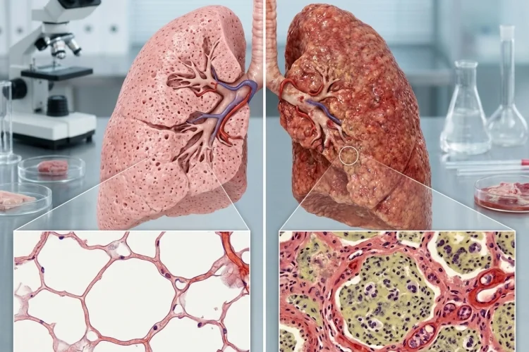
Healthy lungs are mostly about air. With every breath, oxygen travels through branching airways until it reaches millions of tiny air sacs called alveoli. Their thin walls allow oxygen to move into the blood while carbon dioxide travels in the opposite direction.
Pneumonia changes this carefully designed system.
When microorganisms infect the lung, the immune system reacts by producing inflammation. Blood vessels become more active, immune cells move into affected areas, and the normally air-filled alveoli can begin filling with inflammatory fluid, cellular debris, and white blood cells.
The American Lung Association explains that pneumonia causes the alveoli to become inflamed and fill with fluid or pus, making oxygen transfer more difficult.
Understanding how pneumonia changes the air spaces and tissues of the lung helps connect microscopic pathology with familiar symptoms such as cough, fever, shortness of breath, and low oxygen levels.
The basic story is simple: infection reaches the lung, inflammation develops, alveolar spaces fill, gas exchange falls, and the tissue either recovers or develops complications.
What Happens in a Healthy Alveolus?
Before looking at pneumonia, it helps to understand what the lung is supposed to look like.
Alveoli are microscopic air spaces located at the ends of the respiratory tree. Their walls are extremely thin and lie close to networks of pulmonary capillaries. This arrangement minimizes the distance that oxygen and carbon dioxide must travel.
Normally, the inside of an alveolus contains mostly air rather than inflammatory fluid.
The lungs also have several defense mechanisms that prevent microorganisms from reaching these delicate spaces. Mucociliary clearance, antimicrobial substances, airway anatomy, and immune defenses help remove inhaled particles and pathogens before they reach the deeper lung.
Alveolar macrophages provide another important line of defense. These immune cells patrol the air spaces and can destroy many microorganisms without triggering major inflammation.
Pneumonia develops when these defenses are overwhelmed or when a pathogen successfully establishes infection within the lung.
How Infection Reaches the Lung Tissue
Pneumonia can be caused by many different microorganisms, including bacteria, viruses, fungi, and, less commonly, parasites. The CDC defines pneumonia broadly as an infection of the lungs caused by a range of germs.
Microorganisms may enter through inhaled respiratory droplets or through small amounts of material aspirated from the mouth and upper airway.
Once pathogens reach the lower respiratory tract, resident immune cells attempt to eliminate them.
If the infection is too large or the microorganism has effective mechanisms for avoiding host defenses, alveolar macrophages release inflammatory signaling molecules. These signals recruit additional immune cells from the bloodstream.
The immune response is necessary for controlling infection, but it also produces many of the structural changes associated with pneumonia.
The lung is no longer simply dealing with microorganisms. It is now dealing with microorganisms plus inflammation.
Alveoli Begin Filling With Inflammatory Exudate
One of the most important pathological changes in pneumonia occurs inside the alveolar spaces.
Inflammation increases vascular permeability and recruits leukocytes into affected areas. Fluid containing proteins can move from the circulation into lung tissue and alveolar spaces.
In many bacterial pneumonias, neutrophils become prominent inflammatory cells.
Instead of containing mostly air, affected alveoli can become filled with a mixture of neutrophils, protein-rich fluid, fibrin, microorganisms, macrophages, and cellular debris. This material is often called inflammatory exudate.
Imagine replacing the air inside a group of tiny balloons with wet inflammatory material.
Those balloons may still exist structurally, but they can no longer participate effectively in normal ventilation.
This change is central to understanding why pneumonia can make breathing difficult.
Pneumonia Can Produce Lung Consolidation
As increasing numbers of alveoli fill with inflammatory material, affected lung tissue becomes denser.
This process is called consolidation.
Normally, lung tissue feels light and spongy because it contains so much air. Consolidated lung contains much less air because the alveolar spaces have been replaced by fluid and inflammatory cells.
In classic lobar bacterial pneumonia, inflammation can involve a large portion or nearly all of a lung lobe. Pathologically, lobar pneumonia is characterized by acute exudative inflammation and extensive consolidation.
This density change also helps explain why pneumonia can appear as an opacity on a chest X-ray.
Not every pneumonia produces the same distribution, however.
Bronchopneumonia tends to produce patchy areas of inflammation centered around airways, while lobar pneumonia may involve a much larger continuous region.
Other infections, especially many viral infections, may produce more prominent inflammation in the interstitial tissues around alveoli rather than filling the air spaces in exactly the same way.
So pneumonia is not one identical microscopic pattern. The appearance depends partly on the pathogen and the host response.
Why Gas Exchange Becomes More Difficult
The structural changes in pneumonia create an important physiological problem: air and blood are no longer matched effectively.
Blood may still flow through capillaries surrounding infected alveoli. However, if those alveoli are filled with inflammatory exudate instead of fresh air, relatively little oxygen reaches them.
This creates areas where there is perfusion without adequate ventilation.
When enough alveoli are affected, oxygen levels in the blood can fall, producing hypoxemia. Severe pneumonia may therefore cause rapid breathing, shortness of breath, fatigue, confusion, or bluish discoloration associated with inadequate oxygenation.
The problem becomes easier to understand with a simple sequence:
Alveoli fill with inflammatory material → less air enters affected spaces → oxygen transfer decreases → blood oxygen may fall
Pneumonia is therefore not simply an infection sitting inside the lungs. It physically changes the environment where gas exchange is supposed to occur.
What Happens to the Lung Tissue Around the Alveoli?
Pneumonia also affects tissues surrounding the air spaces.
Inflammation can cause pulmonary capillaries to become congested and alveolar walls to appear thickened. In some infections, the inflammatory response involves the interstitium-the supporting tissue between and around alveoli-more prominently than the alveolar spaces themselves.
This matters because gas exchange depends on maintaining a very thin barrier between air and blood.
Swelling, inflammation, and cellular injury can increase the effective distance that oxygen needs to cross.
Different pathogens can also produce different inflammatory patterns. Many typical bacterial pneumonias produce prominent neutrophilic inflammation, whereas some viral infections generate patterns with more lymphocytic or interstitial inflammation.
For pathology students, this is an important reminder: the location and type of inflammatory response provide clues about the disease process.
The Classic Stages of Lobar Pneumonia
Traditional pathology teaching often describes untreated bacterial lobar pneumonia through four overlapping stages.
These stages are useful for understanding how the appearance of lung tissue can change over time, although real patients do not always follow the sequence perfectly.
1. Congestion
Early in the process, blood vessels become congested and the alveoli begin accumulating protein-rich fluid, microorganisms, macrophages, and some neutrophils.
The lung becomes heavy and wet but may not yet be completely consolidated.
2. Red Hepatization
As inflammation intensifies, alveoli become packed with neutrophils, fibrin, fluid, and red blood cells.
The affected tissue becomes firm and takes on a liver-like consistency, which explains the term hepatization.
3. Gray Hepatization
Red blood cells begin breaking down while fibrin and inflammatory cells remain.
The tissue stays firm but develops a more gray appearance.
4. Resolution
If the infection is controlled, inflammatory material is gradually removed. Macrophages help clear debris, while material may also be removed through lymphatic drainage or coughing.
Eventually, surviving lung structures can return toward normal function.
How the Lung Recovers After Pneumonia
One encouraging feature of many pneumonias is that the lung has considerable ability to recover when the infection is successfully controlled.
Neutrophils die or leave the inflammatory site, macrophages remove cellular debris, and excess fluid is cleared.
As the alveolar spaces reopen, ventilation improves and normal gas exchange can gradually return.
Recovery is not always immediate. Clinical symptoms may improve before every inflammatory change in the lung has completely disappeared. The American Lung Association notes that recovery from pneumonia can take weeks in some people.
The final outcome depends on factors such as the microorganism involved, the severity of infection, the patient’s immune response, age, underlying lung disease, and how much tissue injury occurred.
When inflammation resolves effectively, lung architecture may recover remarkably well.
When Pneumonia Causes More Serious Tissue Damage
Most uncomplicated pneumonia does not permanently destroy large amounts of lung tissue, but severe disease can produce significant complications.
Intense infection may cause necrotizing pneumonia, in which areas of lung tissue undergo destruction.
Localized tissue destruction may also result in formation of a lung abscess, a cavity containing necrotic material and pus. Bacterial pneumonia can additionally lead to pleural infection or empyema, sepsis, and respiratory failure.
Severe inflammatory injury may extend beyond localized pneumonia and contribute to acute lung injury.
When damage to the alveolar-capillary barrier becomes widespread, protein-rich fluid can accumulate extensively, making oxygen exchange much more difficult.
Research on pneumonia-associated acute lung injury emphasizes that infection and excessive inflammatory responses can disrupt normal lung homeostasis.
This is why the severity of pneumonia depends on more than the presence of a microorganism.
The degree of inflammation and resulting tissue damage matters too.
A Simple Way to Remember Pneumonia Pathology
For medical learners, pneumonia pathology can be summarized with one chain:
Pathogen enters → alveolar macrophages respond → inflammation develops → neutrophils and fluid enter → alveoli lose air → consolidation develops → gas exchange falls → resolution or complications follow
This framework connects pathology with clinical findings.
A cough helps remove inflammatory secretions. Fever reflects systemic inflammatory signaling. Crackles can occur as air moves through fluid-affected small airways and alveoli. Shortness of breath and low oxygen levels reflect impaired gas exchange.
Instead of memorizing symptoms separately, ask what structural change in the lung could produce each one.
That question turns pneumonia into a logical pathological process.
Pneumonia changes the air spaces and tissues of the lung by turning normally air-filled alveoli into inflamed spaces containing fluid, immune cells, fibrin, microorganisms, and cellular debris.
As more alveoli become involved, lung tissue may consolidate and oxygen transfer into the bloodstream becomes less efficient.
The inflammatory response is essential for eliminating infection, but it is also responsible for many of pneumonia’s symptoms and microscopic changes.
If the infection is controlled, macrophages clear the exudate and alveoli can reopen. Severe disease, however, may produce tissue destruction, abscesses, pleural complications, or respiratory failure.
When studying pneumonia, follow the air spaces first: what normally contains air becomes filled with inflammation-and that single change explains much of the disease.